File:Barr body.JPG
Original file (3,648 × 2,432 pixels, file size: 2.01 MB, MIME type: image/jpeg)
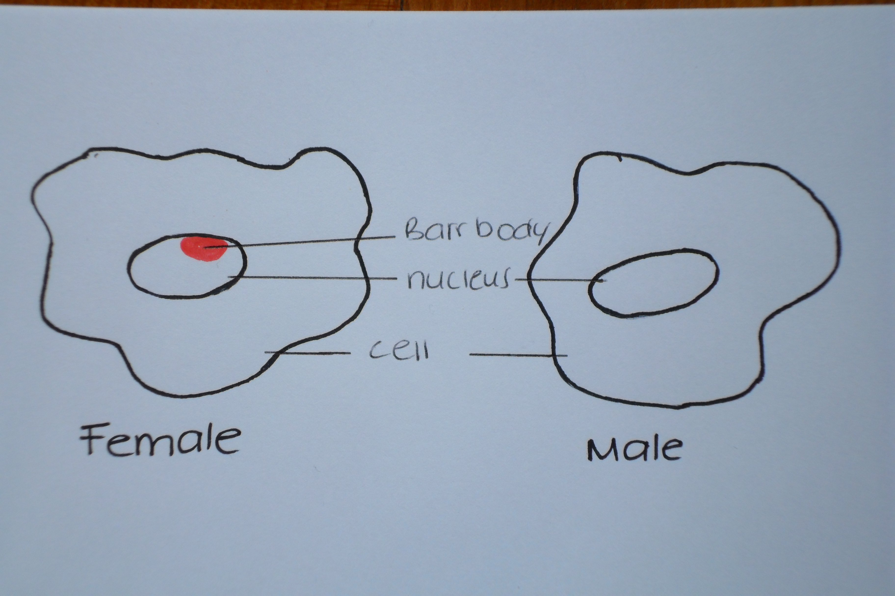
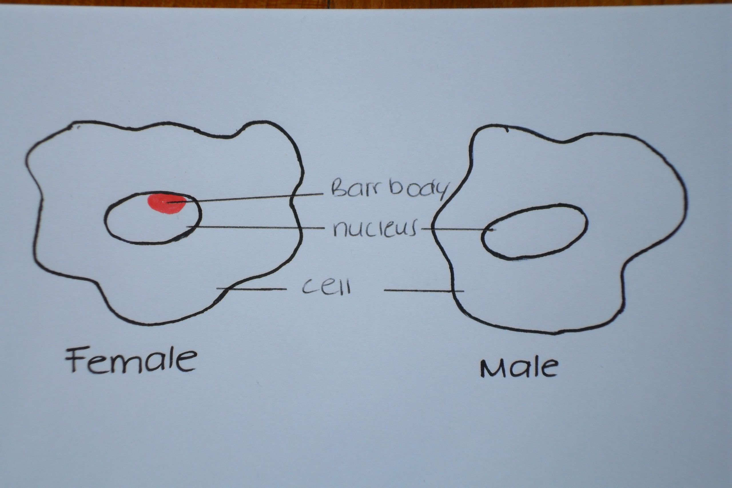
The difference between a female and male cell showing the presence of the Barr or chromatin body discovered by Murray Barr and Ewart Bartram in 1949. This Barr body is able to be seen under the microscope, and therefore the sex of a fetus can be found through analysis of the amniotic fluid.
Illustration by z3292208.
"Beginning six months after publication, I z3292208 grant the public the non-exclusive right to copy, distribute, or display the Work under a Creative Commons Attribution-Noncommercial-Share Alike 3.0 Unported license, as described at http://creativecommons.org/licenses/by-nc-sa/3.0/ and http://creativecommons.org/licenses/by-nc-sa/3.0/legalcode."
File history
Click on a date/time to view the file as it appeared at that time.
| Date/Time | Thumbnail | Dimensions | User | Comment | |
|---|---|---|---|---|---|
| current | 11:45, 11 September 2010 | | 3,648 × 2,432 (2.01 MB) | Z3292208 (talk | contribs) | The difference between a female and male cell showing the presence of the Barr or chromatin body discovered by Murray Barr and Edward Bartram in 1949. This Barr body is able to be seen under the microscope, and therefore the sex of a fetus can be found th |
You cannot overwrite this file.
File usage
The following page uses this file: