File:Bardeen1908a fig02.jpg

Original file (1,000 × 1,295 pixels, file size: 85 KB, MIME type: image/jpeg)
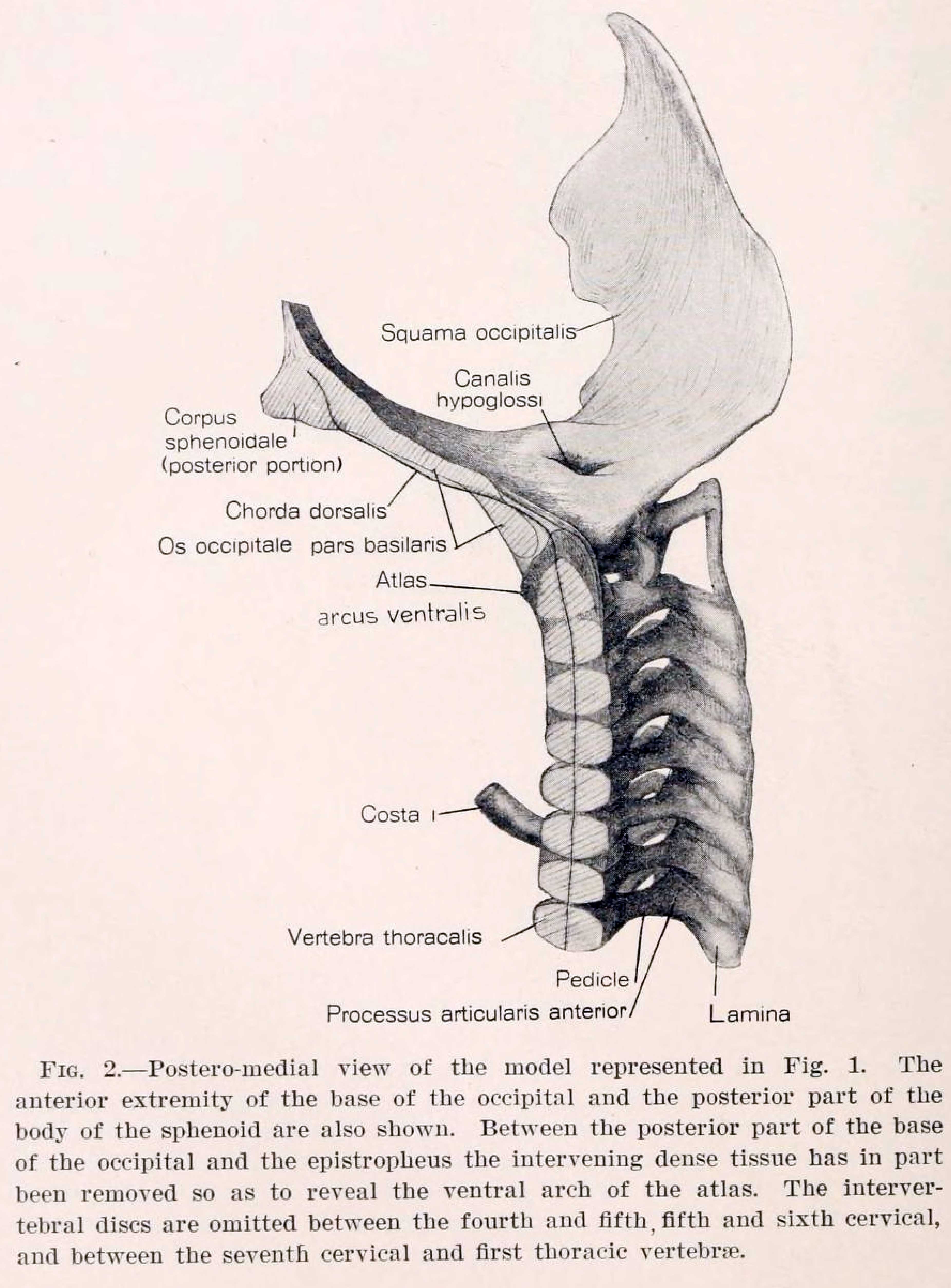
Fig. 2. Postero-medial view of the model represented in Fig 1. The anterior extremity of the base of the occipitnl and the posterior part of the body of the sphenoid are also shown. Between the posterior part of the base of the occipital and the epistropheus the intervening dense tissue has in part been removed so as to reveal the ventral arch of the atlas. The intervertebral discs are omitted between the fourth and fifth fifth and sixth cervical, and between the seventh cervical and first thoracic vertebrae.
Reference
Bardeen CR. Early development of the cervical vertebrae and the base of the occipital bone in man. (1908) Amer. J Anat. 2: 182-186.
Cite this page: Hill, M.A. (2024, May 10) Embryology Bardeen1908a fig02.jpg. Retrieved from https://embryology.med.unsw.edu.au/embryology/index.php/File:Bardeen1908a_fig02.jpg
- © Dr Mark Hill 2024, UNSW Embryology ISBN: 978 0 7334 2609 4 - UNSW CRICOS Provider Code No. 00098G
File history
Click on a date/time to view the file as it appeared at that time.
| Date/Time | Thumbnail | Dimensions | User | Comment | |
|---|---|---|---|---|---|
| current | 16:15, 14 March 2018 | | 1,000 × 1,295 (85 KB) | Z8600021 (talk | contribs) | |
| 16:15, 14 March 2018 | Error creating thumbnail: File with dimensions greater than 12.5 MP | 3,704 × 5,014 (832 KB) | Z8600021 (talk | contribs) | ===Reference=== {{Ref-Bardeen1908a}} {{Footer}} Category:Human Category:VertebraCategory:Axial Skeleton Category:1900'sCategory:Charles Bardeen |
You cannot overwrite this file.
File usage
The following page uses this file: