File:Bailey154.jpg
Original file (898 × 563 pixels, file size: 191 KB, MIME type: image/jpeg)
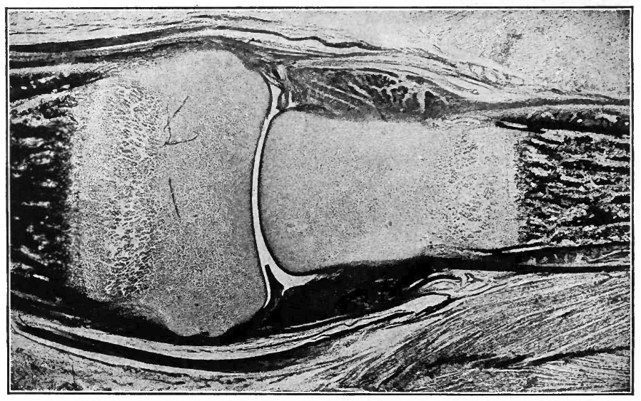
Fig. 154. From longitudinal section of finger of child at birth
Showing developing joint cavity between adjacent ends of phalanges.
The darker portion at each end of the figure indicates the ossification center in the phalanx, the end of the latter (lighter area) being yet cartilaginous.
The dark bands at each side of the joint indicate developing ligaments.
Photograph.
- Text-Book of Embryology: Germ cells | Maturation | Fertilization | Amphioxus | Frog | Chick | Mammalian | External body form | Connective tissues and skeletal | Vascular | Muscular | Alimentary tube and organs | Respiratory | Coelom, Diaphragm and Mesenteries | Urogenital | Integumentary | Nervous System | Special Sense | Foetal Membranes | Teratogenesis | Gallery of All Figures
| Historic Disclaimer - information about historic embryology pages |
|---|
 |
Reference
Bailey FR. and Miller AM. Text-Book of Embryology (1921) New York: William Wood and Co.
Cite this page: Hill, M.A. (2024, May 21) Embryology Bailey154.jpg. Retrieved from https://embryology.med.unsw.edu.au/embryology/index.php/File:Bailey154.jpg
- © Dr Mark Hill 2024, UNSW Embryology ISBN: 978 0 7334 2609 4 - UNSW CRICOS Provider Code No. 00098G
File history
Click on a date/time to view the file as it appeared at that time.
| Date/Time | Thumbnail | Dimensions | User | Comment | |
|---|---|---|---|---|---|
| current | 11:56, 18 January 2011 | | 898 × 563 (191 KB) | S8600021 (talk | contribs) | ==Fig. 154. From longitudinal section of finger of child at birth== Showing developing joint cavity between adjacent ends of phalanges. The darker portion at each end of the figure indicates the ossification center in the phalanx, the end of the latter |
You cannot overwrite this file.
File usage
The following 2 pages use this file: