File:Bailey087.jpg
Original file (928 × 803 pixels, file size: 132 KB, MIME type: image/jpeg)
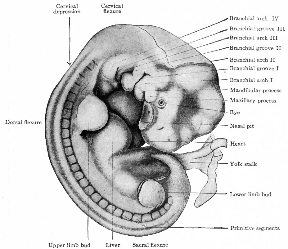
Fig. 87. Human embryo with twenty-seven primitive segments (7 mm, 26 days)
Mall.
In a 7 mm embryo described by Mall (Fig. 87), the flexures are slightly more accentuated than in the 4-mm stage. The branchial arches and grooves are still prominent. The first groove, of which the dorsal part marks the site of the external auditory meatus, is at this time particularly well developed. The eye is a stronger feature than in the preceding stage. The distinct depression in front of the first arch is the nasal fossa. The limb buds are larger than in the 4 mm embryo. The general curvature of the embryo is so sharp at this stage that the rudimentary tail is almost in contact with the head.
Online editor - The term "branchial arch" in human embryos is now more commonly called a "pharyngeal arch".
- Links: Fig. 87 in text | Carnegie stage 14 | Week 5
- Text-Book of Embryology: Germ cells | Maturation | Fertilization | Amphioxus | Frog | Chick | Mammalian | External body form | Connective tissues and skeletal | Vascular | Muscular | Alimentary tube and organs | Respiratory | Coelom, Diaphragm and Mesenteries | Urogenital | Integumentary | Nervous System | Special Sense | Foetal Membranes | Teratogenesis | Gallery of All Figures
| Historic Disclaimer - information about historic embryology pages |
|---|
 |
Reference
Bailey FR. and Miller AM. Text-Book of Embryology (1921) New York: William Wood and Co.
Cite this page: Hill, M.A. (2024, April 27) Embryology Bailey087.jpg. Retrieved from https://embryology.med.unsw.edu.au/embryology/index.php/File:Bailey087.jpg
- © Dr Mark Hill 2024, UNSW Embryology ISBN: 978 0 7334 2609 4 - UNSW CRICOS Provider Code No. 00098G
File history
Click on a date/time to view the file as it appeared at that time.
| Date/Time | Thumbnail | Dimensions | User | Comment | |
|---|---|---|---|---|---|
| current | 15:45, 18 January 2011 | | 928 × 803 (132 KB) | S8600021 (talk | contribs) |
You cannot overwrite this file.