File:Anson1948 fig11.jpg

Original file (1,220 × 981 pixels, file size: 230 KB, MIME type: image/jpeg)
Fig. 11. Reconstruction of the fissular region of the otic capsule in a 180 mm
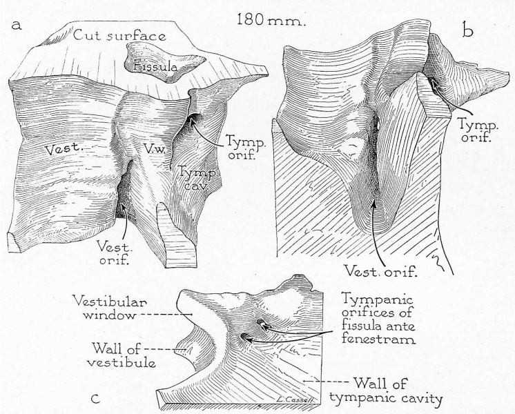
Reconstruction of the fissular region of the otic capsule in a 180 mm. (21 week) fetus (Wisconsin series 45-b) with stapes removed; X 7: (a) viewed from a posterosuperior position, that is, looking toward the anterior wall of the vestibular window and into both orifices of the fissula ante fenestram; (b) viewed from a posteroinferior position, looking upward into the vestibular orifice of the fissula; (c) viewed from a lateral (tyrnpanic) position, looking toward the outer wall of the capsule, where occur, in this specimen, paired tympanic openings of the fissula. Part c is of a block less inclusive than that shown in a and b.
In this specimen the fissula is of aberrant form, being both taller and wider than usual. A cupola-like extension is prolonged cranialward beyond the horizontal level of the tympanic orifice. It contains cartilage (not represented in the reconstruction).
Reference
Anson BJ. and Cauldwell EW. Stapes, fissula ante fenestram and associated structures in man: V . From the fetus of 160 mm to term. (1948) 48(3): 263-300.
Cite this page: Hill, M.A. (2024, May 21) Embryology Anson1948 fig11.jpg. Retrieved from https://embryology.med.unsw.edu.au/embryology/index.php/File:Anson1948_fig11.jpg
- © Dr Mark Hill 2024, UNSW Embryology ISBN: 978 0 7334 2609 4 - UNSW CRICOS Provider Code No. 00098G
File history
Click on a date/time to view the file as it appeared at that time.
| Date/Time | Thumbnail | Dimensions | User | Comment | |
|---|---|---|---|---|---|
| current | 21:07, 16 October 2017 | | 1,220 × 981 (230 KB) | Z8600021 (talk | contribs) | |
| 21:05, 16 October 2017 | 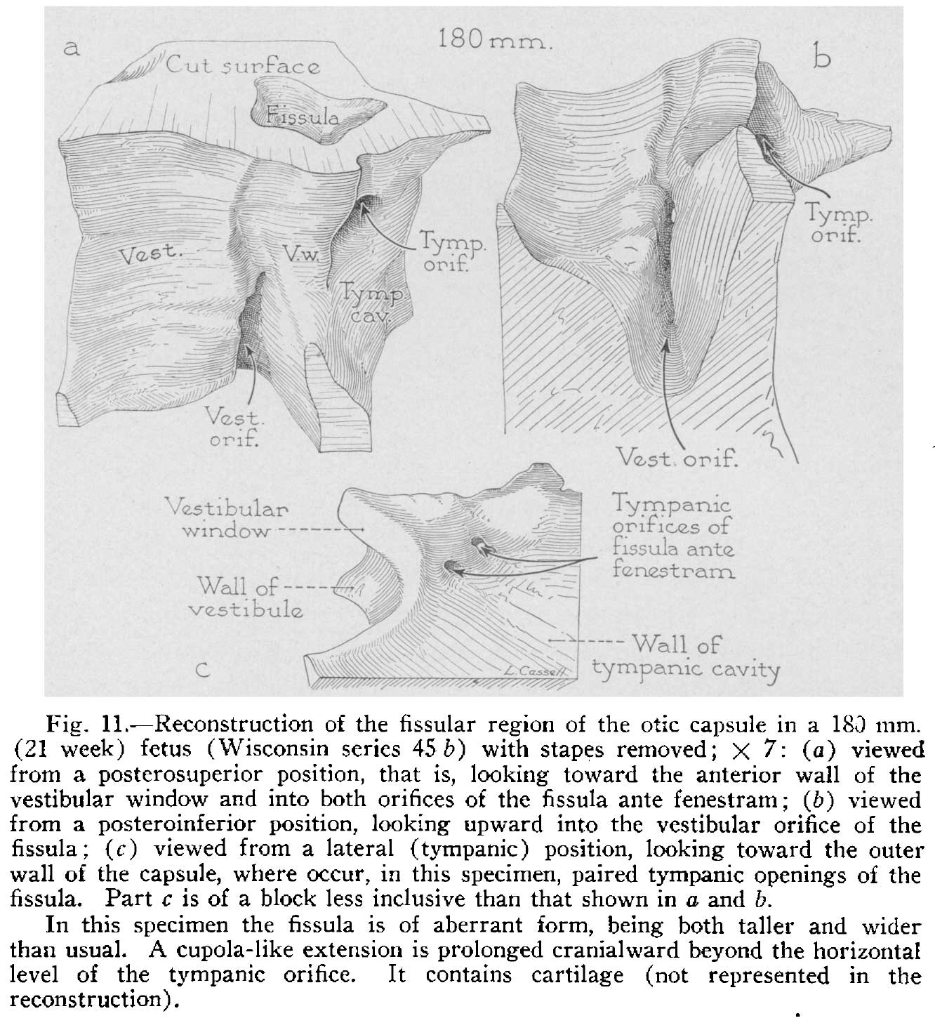 | 1,332 × 1,467 (262 KB) | Z8600021 (talk | contribs) |
You cannot overwrite this file.
File usage
The following 2 pages use this file: