File:Adult epidermis histology 03.jpg
Adult_epidermis_histology_03.jpg (600 × 375 pixels, file size: 46 KB, MIME type: image/jpeg)
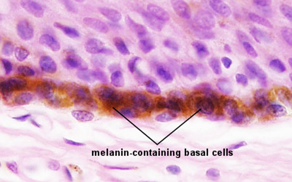
Skin Epithelium Basal Cell Layer
This is an image of the skin epithelial basal cell layer showing melanin (brown) that has been transferred to the keratinocyte stem cells from the nearby melanocytes. Melanin protects the chromosomes of mitotically active basal cells against light-induced damage.
- Integument Histology Links: Adult Skin | Epidermis and Dermis | Thin Skin Epidermis | Thick Skin Epidermis | Elastic Fibres | Basal Cell Melanin | Foundations Practical Support | Integumentary System Development | Histology Stains
Links: Histology | Histology Stains | Blue Histology images copyright Lutz Slomianka 1998-2009. The literary and artistic works on the original Blue Histology website may be reproduced, adapted, published and distributed for non-commercial purposes. See also the page Histology Stains.
Cite this page: Hill, M.A. (2024, April 28) Embryology Adult epidermis histology 03.jpg. Retrieved from https://embryology.med.unsw.edu.au/embryology/index.php/File:Adult_epidermis_histology_03.jpg
- © Dr Mark Hill 2024, UNSW Embryology ISBN: 978 0 7334 2609 4 - UNSW CRICOS Provider Code No. 00098G
File history
Click on a date/time to view the file as it appeared at that time.
| Date/Time | Thumbnail | Dimensions | User | Comment | |
|---|---|---|---|---|---|
| current | 13:26, 26 March 2012 | | 600 × 375 (46 KB) | Z8600021 (talk | contribs) |
You cannot overwrite this file.
File usage
The following 5 pages use this file: