File:Meckel.jpg
From Embryology

Size of this preview: 719 × 599 pixels.
Original file (800 × 667 pixels, file size: 181 KB, MIME type: image/jpeg)
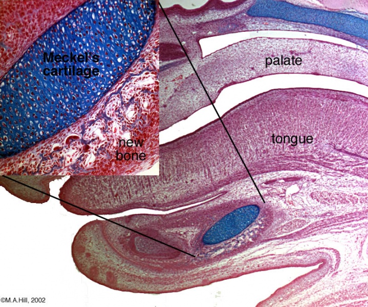
Meckel's Cartilage
Head histology showing detailed view of Meckel's cartilage from the first pharyngeal arch. Inset shows detail of both cartilage and bone development.
- Meckel's cartilage - is not being replaced by bone, as in endochondral ossification, but it will later degenerate after the bony mandible forms.
- Mandible bone - intramembranous ossification occurring beside the original cartilage model.
Reference
Image Source: UNSW Embryology, no reproduction without permission.
Cite this page: Hill, M.A. (2024, May 15) Embryology Meckel.jpg. Retrieved from https://embryology.med.unsw.edu.au/embryology/index.php/File:Meckel.jpg
- © Dr Mark Hill 2024, UNSW Embryology ISBN: 978 0 7334 2609 4 - UNSW CRICOS Provider Code No. 00098G
File history
Click on a date/time to view the file as it appeared at that time.
| Date/Time | Thumbnail | Dimensions | User | Comment | |
|---|---|---|---|---|---|
| current | 11:15, 31 August 2009 | | 800 × 667 (181 KB) | S8600021 (talk | contribs) | Meckel's cartilage of the first pharyngeal arch. http://embryology.med.unsw.edu.au/Notes/head6.htm |
You cannot overwrite this file.
File usage
The following 15 pages use this file:
- 2009 Lecture 11
- 2010 Lecture 11
- AACP Meeting 2013 - Face Embryology
- ANAT2241 Bone, Bone Formation and Joints
- Abnormal Development - Cleft Palate
- BGD Lecture - Face and Ear Development
- Bone Development
- Bone Histology
- Cartilage Histology
- Embryology History - Johann Meckel
- Head Development
- Lecture - Head Development
- M
- Musculoskeletal System - Bone Development
- Palate Development