Category:Head
From Embryology
This Embryology category shows media and pages related tho head development. This can include pharyngeal arches, skull, oral cavity, nasal cavity, upper gastrointestinal tract, upper respiratory tract, endocrine, vascular, lymphatic and neural development features. Each of these topics is also covered by specific notes.
Subcategories
This category has the following 8 subcategories, out of 8 total.
Pages in category 'Head'
The following 189 pages are in this category, out of 189 total.
2
A
B
- BGD Lecture - Face and Ear Development
- BGD Practical - Face and Ear Quiz
- BGD Practical - Face Quiz
- Template:BGDB Face
- BGDB Face and Ear - Abnormalities
- BGDB Face and Ear - Early Embryo
- BGDB Face and Ear - Fetal
- BGDB Face and Ear - Late Embryo
- BGDB Face and Ear - Postnatal
- BGDB Face and Ear - Trilaminar Embryo
- BGDB Practical - Face and Ear Development
- Talk:BGDB Practical - Face and Ear Development
- BGDB Practical - Face and Ear Development Interactive
- Template:BGDB Practical 6 - Abnormalities Interactive
- Template talk:BGDB Practical 6 - Abnormalities Interactive
- Template:BGDB Practical 6 - Early Embryo Interactive
- Template:BGDB Practical 6 - Fetal Interactive
- Template:BGDB Practical 6 - Late Embryo Interactive
- Template:BGDB Practical 6 - Postnatal Interactive
- Template:BGDB Practical 6 - Trilaminar Embryo Interactive
- Book - Contributions to Embryology Carnegie Institution No.48
- Book - Contributions to Embryology Carnegie Institution No.55
- Book - Human Embryology and Morphology 1
- Book - Text-Book of Embryology 8
- Buccopharyngeal membrane
- Template:Buccopharyngeal membrane
F
H
- Template:Head
- Template:Head abnormalities
- Template:Head and Neck Links
- Head Development
- Head Development - Abnormalities
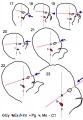
- Head Development - Carnegie Stage 22
- Template:Head Links
- Template:Holoprosencephaly
- Human Embryology and Morphology 12
- Human Embryology and Morphology 17
- Template:Human Eyelid timeline table
- Human System Development
M
P
- Template:Palate
- Palate Development
- Palate Development 1 Movie
- Palate Development 2 Movie
- Paper - A human foetus exhibiting iniencephaly and other abnormalities (1922)
- Paper - An anencephalic embryo of 25 mm CRL
- Paper - Contribution to the structure and development of the vertebrate head
- Paper - Contribution to the structure and development of the vertebrate head 1
- Paper - Contribution to the structure and development of the vertebrate head 2
- Paper - Contribution to the structure and development of the vertebrate head 3
- Paper - Description of a reconstruction of the head of a thirty-millimetre embryo (1910)
- Paper - Development of olfactory and related structures in staged human embryos
- Paper - Evolutionary factors in the production of pharyngeal diverticula
- Paper - Extroversion of the cerebral hemispheres in a human embryo (1934)
- Paper - Malformations of the human body from a new point of view 1+2
- Paper - Normal facial growth in children (1937)
- Paper - Observations on metopism (1917)
- Paper - On the premature obliteration of sutures in the human skull (1915)
- Paper - On the presence of a series of ectodermal placodes in the head region of a sparrow embryo (1928)
- Paper - On the relation of the chorda dorsalis to the anlage of the pharyngeal bursa or median pharyngeal recess (1912)
- Paper - On the relation of the head chorda to the pharyngeal epithelium in the pig embryo
- Paper - On the so-called ultimobranchial body of the mammalian embryo (1915)
- Paper - Pouches of the pharynx and oesophagus with special reference to the embryological and morphological aspects
- Paper - Preliminary note on the skull of a human fetus of 43 mm greatest length
- Paper - Primary neuromeres and head segmentation (1922)
- Paper - The Anatomy of the Head End of a 20 mm Human Embryo
- Paper - The aortic arch derivatives in human adult (1951)
- Paper - The cartilaginous skull of a human embryo twenty-one millimeters in length (1920)
- Paper - The chondrocranium of a 20 mm human embryo
- Paper - The development of the anterior post-otic somites in the rabbit
- Paper - The development of the cerebrospinal fluid spaces and choroid plexuses in the chick (1937)
- Paper - The development of the cranial arteries in the human embryo
- Paper - The development of the first branchial arch in man and the fate of Meckel's cartilage
- Paper - The development of the human chin (1917)
- Paper - The Development of the Human Mandibular Joint
- Paper - The development of the human pharynx
- Paper - The Development of the Nose and of the Pharynx and its Derivatives in Man
- Paper - The development of the sphenoidal sinus in man and its homology in mammals (1927)
- Paper - The development of the subcutaneous vascular plexus in the head of the human embryo (1923)
- Paper - The developmental alterations in the vascular system of the brain of the human embryo (1921)
- Paper - The genesis, development, and adult anatomy of the nasofrontal region in man
- Paper - The lateral wall of the cavum nasi in man, with especial reference to the various developmental stages
- Paper - The Long Fox lecture - The development of the human skull (1910)
- Paper - The pharyngeal pouches and their derivatives in the mammalia
- Paper - The prenatal development of the human temporomandibular joint
- Paper - The primordial cranium of erinaceus europaeus (1918)
- Paper - The primordial cranium of Erinaceus europaeus (1918)
- Paper - The primordial cranium of microtus amphibius (water-rat), as determined by sections and a model of the 25-mm stage (1917)
- Paper - The primordial cranium of miniopterus schreibersi at the 17 millimetre total length stage (1919)
- Paper - The second visceral arch and groove in the tubo-tympanic region
- Paper - The sinus maxillaris and its relations in the embryo, child, and adult man
- Paper - Three demonstrations on congenital melformations of palate, face, and neck
- Paper - Transformation of the aortic-arch system during the development of the human embryo (1922)
- Paper - Vertebrate cephalogenesis 1 (1890)
- Paper - Vertebrate cephalogenesis 2 (1892)
- Paper - Vertebrate cephalogenesis 4 (1919)
- Paper The development of the subcutaneous vascular plexus in the head of the human embryo (1923)
- Template:Pharyngeal arch
- Template:Pharyngeal Arch table
R
- Template:Ref-Allis1938
- Template:Ref-Ayers1890
- Template:Ref-Ayers1892
- Template:Ref-Bartelmez1924
- Template:Ref-Boulgakow1926
- Template:Ref-Dickie1914
- Template:Ref-Fawcett1910head
- Template:Ref-Fawcett1911
- Template:Ref-Finley1922
- Template:Ref-Finley1923
- Template:Ref-Huber1912
- Template:Ref-JacksonAJ1935
- Template:Ref-Keith1909
- Template:Ref-Keith1932a
- Template:Ref-Keith1932b
- Template:Ref-Kingsbury1915a
- Template:Ref-Kingsbury1915b
- Template:Ref-Locy1895
- Template:Ref-Mann1921
- Template:Ref-Moffatt1957
- Template:Ref-Padget1956
- Template:Ref-Rand1917
- Template:Ref-Raven1933
- Template:Ref-Schaeffer1910
- Template:Ref-Schaeffer1916
- Template:Ref-Stunkard1922
- Template:Ref-Wallis1917
- Template:Ref-Young1937
- Template:Reichert’s cartilage
S
Media in category 'Head'
The following 200 files are in this category, out of 308 total.
(previous page) (next page) Bailey096.jpg 693 × 501; 59 KB
 Bailey097.jpg 776 × 674; 71 KB
 Bailey098.jpg 680 × 432; 47 KB
 Bailey099.jpg 704 × 464; 52 KB
 Bailey132+133.jpg 940 × 570; 101 KB
 Bailey132.jpg 466 × 413; 43 KB
 Bailey133.jpg 806 × 655; 85 KB
 Bailey135.jpg 940 × 965; 216 KB
 Bailey136.jpg 835 × 566; 114 KB
 Bailey137.jpg 672 × 539; 73 KB
 Bailey138.jpg 831 × 400; 62 KB
 Bailey139.jpg 961 × 671; 96 KB
 Bailey140.jpg 793 × 505; 58 KB
 Bailey141.jpg 761 × 323; 66 KB
 Bailey142.jpg 778 × 479; 72 KB
 Bailey191.jpg 863 × 509; 109 KB
 Bailey192.jpg 960 × 806; 133 KB
 Bailey193.jpg 747 × 848; 94 KB
 Baileytable02.jpg 884 × 1,109; 182 KB
 Bat-craniofacial development.jpg 600 × 720; 140 KB
 BGDB PracManual 2011 Practical 6.pdf ; 426 KB
 Bilateral cleft palate.jpg 214 × 300; 11 KB
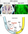
 Chicken HH20 MyoR expression 01.jpg 960 × 917; 121 KB
 Chicken HH20 MyoR expression 02.jpg 1,200 × 620; 167 KB
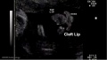
 Cleft lip 01.jpg 585 × 438; 34 KB
 Cleft lip 02.jpg 641 × 362; 22 KB
 Cleft palate.jpg 653 × 776; 124 KB
 Cranial neural crest skeletal fate 01.jpg 800 × 633; 59 KB
 Cytomegalovirus induced micrognathia and abnormal skeletogenesis.jpg 1,200 × 1,105; 355 KB
 Dickie1914 fig01.jpg 463 × 611; 57 KB
 Dickie1914 fig02.jpg 443 × 526; 58 KB
 Dickie1914 fig03.jpg 672 × 516; 64 KB
 Dickie1914 fig04.jpg 755 × 578; 71 KB
 Dickie1914 fig05.jpg 613 × 567; 51 KB
 Dickie1914 fig06.jpg 504 × 607; 57 KB
 Dickie1914 fig07.jpg 407 × 577; 74 KB
 Dickie1914 fig08.jpg 538 × 523; 93 KB
 Dickie1914 fig09.jpg 351 × 526; 31 KB
 Dickie1914 fig10.jpg 337 × 525; 33 KB
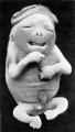
 Fetal facial expression 01.jpg 1,200 × 1,094; 122 KB
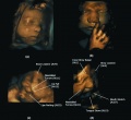 Fetal facial expression 02.jpg 1,914 × 1,762; 205 KB
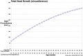 Fetal head growth circumference graph01.jpg 905 × 613; 58 KB
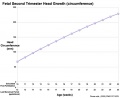 Fetal head growth circumference graph02.jpg 800 × 650; 44 KB
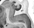
 Fetal head lateral.jpg 632 × 447; 34 KB
 Fetal head medial.jpg 632 × 447; 34 KB
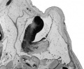
 Fetal head section 01.jpg 1,200 × 821; 186 KB
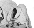
 Fetal head section.jpg 1,200 × 821; 167 KB
 Fetal mandible and lower lip 01.jpg 1,028 × 557; 57 KB
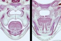
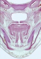
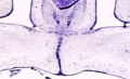
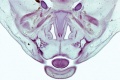 Fetal week 10 hard palate 01.jpg 800 × 532; 77 KB
 Fetal week 10 hard palate 02.jpg 398 × 633; 66 KB
 Fetal week 10 hard palate 03.jpg 600 × 450; 122 KB
 Fetal week 10 hard palate 04.jpg 1,198 × 795; 196 KB
 Fetal week 10 hard palate 06.jpg 534 × 778; 88 KB
 Fetal week 10 hard palate 07.jpg 534 × 778; 97 KB
 Fetal week 10 palate 01.gif 534 × 778; 1.14 MB
 Fetal week 10 palate 01.mp4 ; 427 KB
 Fetal week 10 palate icon.jpg 534 × 778; 100 KB
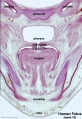
 Fetal week 10 soft palate 01.jpg 571 × 784; 95 KB
 Fetal week 10 soft palate 02.jpg 534 × 778; 87 KB
 Fetal week 10 soft palate 03.jpg 534 × 778; 95 KB
 Fetal week 14 head bone lateral 01.jpg 1,000 × 773; 107 KB
 Fetal week 9 hard palate fusion 01.jpg 661 × 400; 51 KB
 Fetal week 9 head lateral 01.jpg 700 × 600; 78 KB
 Finley1923 fig01.jpg 494 × 968; 53 KB
 Finley1923 fig02.jpg 700 × 800; 77 KB
 Finley1923 fig03.jpg 600 × 512; 56 KB
 Finley1923 fig04.jpg 700 × 627; 61 KB
 Finley1923 fig05.jpg 674 × 800; 146 KB
 Finley1923 fig06.jpg 729 × 800; 95 KB
 Finley1923 fig07.jpg 314 × 800; 40 KB
 Finley1923 fig08.jpg 296 × 800; 32 KB
 Finley1923 fig09.jpg 456 × 800; 49 KB
 Finley1923 fig10.jpg 221 × 802; 17 KB
 Finley1923 fig11.jpg 432 × 800; 38 KB
 Finley1923 fig12.jpg 593 × 800; 48 KB
 Finley1923 fig13.jpg 594 × 800; 51 KB
 Finley1923 Plate 1.jpg 776 × 1,000; 151 KB
 Finley1923 Plate 2.jpg 864 × 1,200; 153 KB
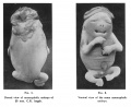
 Foster113.jpg 927 × 512; 57 KB
 Frazer1926 fig01.jpg 1,200 × 804; 137 KB
 Frazer1926 fig02.jpg 991 × 833; 145 KB
 Frazer1926 fig03.jpg 1,200 × 804; 69 KB
 Frazer1926 fig04.jpg 1,200 × 804; 95 KB
 Frazer1926 fig05.jpg 1,000 × 643; 85 KB
 Frazer1926 fig06.jpg 563 × 811; 35 KB
 Frazer1926 fig07.gif 554 × 600; 134 KB
 Frazer1926 fig07.jpg 1,229 × 996; 95 KB
 Frazer1926 fig08.jpg 616 × 789; 38 KB
 Frazer1926 plate01.jpg 1,914 × 2,681; 469 KB
 Gray0043.jpg 800 × 496; 50 KB
 Gray0048.jpg 500 × 429; 30 KB
 Gray0050.jpg 600 × 332; 46 KB
 Gray0051.jpg 600 × 423; 91 KB
 Gray0379.jpg 713 × 600; 114 KB
 Gray0593.jpg 550 × 437; 66 KB
 Gray0603.jpg 800 × 480; 69 KB
 Gray0604.jpg 716 × 500; 65 KB
 Gray0605.jpg 614 × 600; 98 KB
 Gray0947.jpg 600 × 398; 56 KB
 Gray0994.jpg 600 × 861; 151 KB
 Gray1013.jpg 562 × 500; 56 KB
 Gray1014.jpg 619 × 600; 95 KB
 Gray1015.jpg 598 × 300; 62 KB
 Gray1016.jpg 252 × 400; 18 KB
 Gray1017.jpg 450 × 334; 31 KB
 Gray1018.jpg 390 × 400; 53 KB
 Gray1019.jpg 644 × 650; 87 KB
 Gray1020.jpg 667 × 400; 53 KB
 Gray1022.jpg 629 × 400; 45 KB
 Gray1023.jpg 585 × 400; 47 KB
 Gray1024.jpg 800 × 691; 117 KB
 Gray1027.jpg 598 × 600; 101 KB
 Gray1029.jpg 700 × 435; 75 KB
 Gray1030.jpg 481 × 800; 122 KB
 Gray1202.jpg 563 × 500; 63 KB
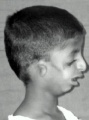 Head - Treacher Collins Syndrome 01.jpg 291 × 391; 16 KB
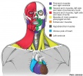 Head and heart muscle cartoon.jpg 874 × 800; 129 KB
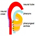 Head arches cartoon.jpg 394 × 402; 29 KB
 Hertwig286.jpg 642 × 800; 126 KB
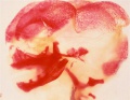
 Human embryo head week 6 to 8.jpg 540 × 780; 66 KB
 Human fetal temporal bone and mandible 01.jpg 1,200 × 805; 170 KB
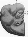
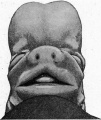
 Human holoprosencephaly cyclopia dissection.jpg 600 × 340; 37 KB
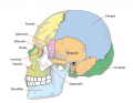 Human skull lateral simplified.png 740 × 576; 138 KB
 Human stage16 face 01.jpg 500 × 504; 20 KB
 Human stage17 face 01.jpg 500 × 504; 21 KB
 Human stage18 face 01.jpg 500 × 504; 23 KB
 Human- fetal week 10 cerebellum A.jpg 347 × 284; 24 KB
 Human- fetal week 10 cerebellum B.jpg 347 × 284; 21 KB
 Human- fetal week 10 cerebellum C.jpg 347 × 284; 25 KB
 Human- fetal week 10 cerebellum D.jpg 347 × 284; 23 KB
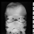
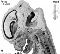 Human- fetal week 10 head A.jpg 600 × 544; 113 KB
 Human- fetal week 10 head A1.jpg 1,200 × 1,088; 159 KB
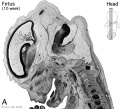
 Human- fetal week 10 head B.jpg 600 × 544; 66 KB
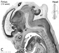 Human- fetal week 10 head C.jpg 600 × 544; 118 KB
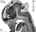 Human- fetal week 10 head D.jpg 600 × 544; 111 KB
 Hunter1934 fig01-02.jpg 1,594 × 1,308; 188 KB
 Hunter1934 fig01.jpg 600 × 1,062; 64 KB
 Hunter1934 fig02.jpg 565 × 1,000; 60 KB
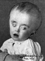 Hydrocephalus.jpg 320 × 432; 29 KB
 Keibel Mall 066-071.jpg 610 × 800; 58 KB
 Keibel Mall 068-069.jpg 1,000 × 358; 35 KB
 Keibel Mall 070-071.jpg 1,000 × 490; 45 KB
 Keibel Mall 2 257.jpg 869 × 800; 120 KB
 Keibel Mall 2 258.jpg 927 × 540; 78 KB
 Keibel Mall 2 334.jpg 1,000 × 332; 50 KB
 Keibel Mall 2 335.jpg 793 × 800; 61 KB
 Keibel Mall 2 336.jpg 777 × 800; 34 KB
 Keibel Mall 231.jpg 450 × 511; 21 KB
 Keibel Mall 232.jpg 450 × 511; 35 KB
 Keibel Mall 266.jpg 740 × 608; 77 KB
 Keibel Mall 367.jpg 857 × 850; 112 KB
 Keibel Mall 368.jpg 800 × 599; 63 KB
 Keibel Mall 370.jpg 900 × 740; 111 KB
 Keith1902 fig001.jpg 823 × 750; 75 KB
 Keith1902 fig002.jpg 932 × 800; 69 KB
 Keith1902 fig003.jpg 1,000 × 700; 113 KB
 Keith1902 fig004.jpg 580 × 314; 27 KB
 Keith1902 fig005.jpg 650 × 557; 36 KB
 Keith1902 fig006.jpg 1,000 × 689; 112 KB
 Keith1902 fig007.jpg 946 × 700; 80 KB
 Keith1902 fig008.jpg 925 × 700; 122 KB
 Keith1902 fig009.jpg 1,000 × 568; 78 KB
 Keith1902 fig010a-c.jpg 800 × 1,000; 112 KB
 Keith1902 fig010d.jpg 800 × 581; 66 KB
 Keith1902 fig011.jpg 954 × 500; 66 KB
 Keith1902 fig012.jpg 875 × 500; 75 KB
 Keith1902 fig013.jpg 662 × 800; 67 KB
 Keith1902 fig014.jpg 707 × 1,000; 98 KB
 Keith1902 fig015a.jpg 971 × 600; 74 KB
 Keith1902 fig015b.jpg 1,000 × 719; 97 KB
 Keith1902 fig021a.jpg 923 × 700; 0 bytes
 Keith1902 fig021b.jpg 800 × 727; 75 KB
 Keith1902 fig022.jpg 807 × 800; 92 KB
 Keith1902 fig023.jpg 800 × 411; 85 KB
 Keith1902 fig024.jpg 600 × 433; 47 KB
 Keith1902 fig025.jpg 876 × 800; 61 KB
 Keith1902 fig026.jpg 1,000 × 728; 92 KB
 Keith1902 fig027.jpg 881 × 800; 93 KB
 Keith1902 fig028.jpg 707 × 800; 77 KB
 Keith1902 fig029.jpg 872 × 600; 81 KB
 Keith1902 fig030.jpg 803 × 700; 87 KB
 Keith1902 fig031.jpg 1,000 × 557; 151 KB
 Keith1902 fig032.jpg 1,000 × 496; 81 KB
 Keith1902 fig033.jpg 971 × 800; 139 KB
 Keith1902 fig034.jpg 921 × 700; 110 KB
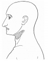
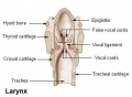 Larynx.jpg 473 × 345; 16 KB
 Lizard embryo 11.jpg 1,200 × 900; 122 KB
 Low 14.jpg 508 × 554; 60 KB
 Mall1906 fig04.jpg 1,114 × 1,044; 137 KB
 ME16 001.jpg 1,740 × 2,500; 557 KB
 ME16 002.jpg 1,037 × 1,500; 272 KB
 ME18 001.jpg 773 × 1,200; 176 KB
 Meckel.jpg 800 × 667; 181 KB
 Monosomy 18p syndrome facial features.jpg 967 × 727; 106 KB
 Mouse Bmp4 expression face 01.jpg 1,200 × 322; 58 KB
 Mouse Bmp4 expression limb and face 01.jpg 1,200 × 513; 91 KB
- Mouse face Bmp4.mp4 ; 480 KB
 Mouse head E11.5 microCT 01.jpg 1,409 × 1,200; 308 KB
 Mouse head TFII-I expression 01.jpg 1,000 × 862; 298 KB
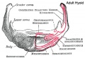 Musculoskeletal- adult hyoid.jpg 450 × 309; 45 KB


