File:Human- fetal week 10 head D.jpg
From Embryology
No higher resolution available.
Human-_fetal_week_10_head_D.jpg (600 × 544 pixels, file size: 111 KB, MIME type: image/jpeg)
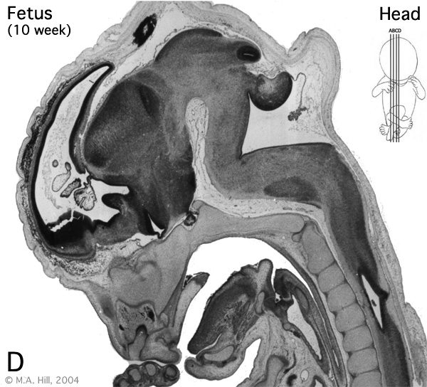
Human Fetus (week 10) Fetal
female, 10 week, 40 mm CRL, early fetal, sagittal section, pelvic region
This stage of development is after the embryonic period (up to week 8) but still only 2 weeks into early fetal development.
Section D is the most midline of all sections. Planes A, B, C and D move towards the midline.
Original file name: H10wkHeadD.jpg
Related Images
Fetus (week 10) Planes A (most lateral), B (lateral), C (medial) and D (midline) from lateral towards the midline.
- Human Fetus - most lateral | lateral | medial | midline
- Head - most lateral | lateral | medial | midline
- Cerebellum - most lateral | lateral | medial | midline
- Urogenital Unlabelled - most lateral | lateral | medial | midline
- Urogenital Labelled - most lateral | lateral | medial | midline
- Large Images - midline
- Image Source: UNSW Embryology, no reproduction without permission.
File history
Click on a date/time to view the file as it appeared at that time.
| Date/Time | Thumbnail | Dimensions | User | Comment | |
|---|---|---|---|---|---|
| current | 14:23, 27 April 2010 | | 600 × 544 (111 KB) | S8600021 (talk | contribs) |
You cannot overwrite this file.