File:Whitehead1904 fig05.jpg
From Embryology

Size of this preview: 667 × 599 pixels.
Original file (800 × 719 pixels, file size: 124 KB, MIME type: image/jpeg)
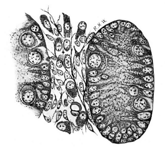
Fig. 5. Pig Embryo 1.5 cm
The tubules are much larger, the spaces and the Leydig’s cells much smaller than in preceding stages. Mallory‘s connective-tlssue stain. X 800.
| Historic Disclaimer - information about historic embryology pages |
|---|
 |
- Links: fig 1 | fig 2 | fig 3 | fig 4 | fig 5 | fig 6 | fig 7 | fig 8 | fig 9 | fig 10 | 1904 Whitehead | Historic Embryology Papers | Testis Development
Reference
Whitehead RH. The embryonic development of the interstitial cells of Leydig. (1904) Amer. J Anat. 3:167-182.
Cite this page: Hill, M.A. (2024, May 8) Embryology Whitehead1904 fig05.jpg. Retrieved from https://embryology.med.unsw.edu.au/embryology/index.php/File:Whitehead1904_fig05.jpg
- © Dr Mark Hill 2024, UNSW Embryology ISBN: 978 0 7334 2609 4 - UNSW CRICOS Provider Code No. 00098G
File history
Click on a date/time to view the file as it appeared at that time.
| Date/Time | Thumbnail | Dimensions | User | Comment | |
|---|---|---|---|---|---|
| current | 09:30, 24 January 2017 | | 800 × 719 (124 KB) | Z8600021 (talk | contribs) | |
| 09:29, 24 January 2017 | 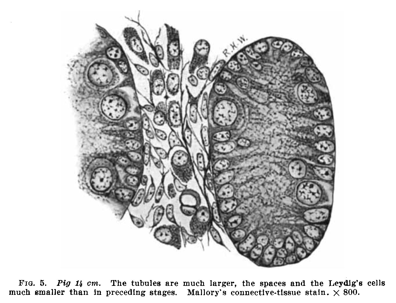 | 1,361 × 1,043 (215 KB) | Z8600021 (talk | contribs) | {{Whitehead1904 figures}} |
You cannot overwrite this file.
File usage
The following page uses this file: