File:Whillis1940 plate03.jpg

Original file (1,280 × 1,419 pixels, file size: 259 KB, MIME type: image/jpeg)
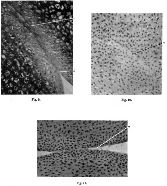
Plate III
Fig. 9. Section of elbow of newly born rat. (Thionin.) X340. a, continuity of matrix across joint line; b, liquefaction of matrix at edge near synovial cavity.
Fig. 10. Section of elbow of newly born rat, nearer the edge of the bond of union than fig. 9. x 340. a, degenerating matrix.
Fig. 11. The process of separation almost complewd. x 340. a, liquefaction of the matrix.
Reference
Whillis J. The development of synovial joints. (1940) J Anat. 74(Pt 2): 277-283. PMID: 17104813
Cite this page: Hill, M.A. (2024, May 11) Embryology Whillis1940 plate03.jpg. Retrieved from https://embryology.med.unsw.edu.au/embryology/index.php/File:Whillis1940_plate03.jpg
- © Dr Mark Hill 2024, UNSW Embryology ISBN: 978 0 7334 2609 4 - UNSW CRICOS Provider Code No. 00098G
File history
Click on a date/time to view the file as it appeared at that time.
| Date/Time | Thumbnail | Dimensions | User | Comment | |
|---|---|---|---|---|---|
| current | 18:47, 16 August 2017 | | 1,280 × 1,419 (259 KB) | Z8600021 (talk | contribs) | |
| 18:46, 16 August 2017 |  | 1,884 × 2,507 (765 KB) | Z8600021 (talk | contribs) |
You cannot overwrite this file.
File usage
The following page uses this file: