File:Ultrasound12wk 3D.jpg
From Embryology
No higher resolution available.
Ultrasound12wk_3D.jpg (512 × 398 pixels, file size: 18 KB, MIME type: image/jpeg)
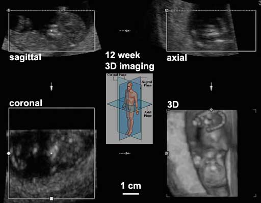
Ultrasound image 12 week Fetus showing Three Dimensional (3d) Axes

Anatomical planes are used in gross anatomy, clinical imaging and histology to define the direction of viewing in the standard anatomical position.
- coronal plane - (frontal plane) is any vertical plane that divides the body into ventral and dorsal sections.
- sagittal plane - vertical plane which passes from anterior to posterior, dividing the body into right and left halves. Midsagittal (median) plane is located in the midline, all other planes are parasagittal planes.
- transverse plane - (cross-section, horizontal plane, axial plane, or transaxial plane) is plane that divides the body into superior and inferior parts and is perpendicular to the coronal and sagittal planes.
File history
Click on a date/time to view the file as it appeared at that time.
| Date/Time | Thumbnail | Dimensions | User | Comment | |
|---|---|---|---|---|---|
| current | 16:31, 5 August 2009 | | 512 × 398 (18 KB) | S8600021 (talk | contribs) | Ultrasound image 12 week fetus, showing 3 dimensional (3d) axes Original file name: 12wk2_3D.jpg Image source: UNSW Embryology http://embryology.med.unsw.edu.au/Movies/usound/Hum3D.htm |
You cannot overwrite this file.
File usage
The following 13 pages use this file:
- 2010 BGD Tutorial - Applied Embryology and Teratology
- 2010 Foundations Lecture - Introduction to Human Development
- BGD Tutorial - Applied Embryology and Teratology
- Fetal Surgery
- Foundations Lecture - Introduction to Human Development
- Foundations Practical - Additional Resources
- Neonatal Diagnosis
- Pre-Medicine Program - Embryology
- Prenatal Diagnosis
- Prenatal Genetic Diagnosis
- U
- Ultrasound
- Ultrasound - Nuchal Translucency