Fig. 8. Human ovum in discus proligerus, shows first polar body undergoing division in the ovum in which the second polar body is in process of being extruded
x 900.
| Historic Disclaimer - information about historic embryology pages
|
| Pages where the terms "Historic" (textbooks, papers, people, recommendations) appear on this site, and sections within pages where this disclaimer appears, indicate that the content and scientific understanding are specific to the time of publication. This means that while some scientific descriptions are still accurate, the terminology and interpretation of the developmental mechanisms reflect the understanding at the time of original publication and those of the preceding periods, these terms, interpretations and recommendations may not reflect our current scientific understanding. (More? Embryology History | Historic Embryology Papers)
|
- Human Ovum Links: Fig 1. Prophase I | Fig 2. | | Fig 3. | Plate 10 | Plate 11 | Plate 12
| Online Editor Notes
|
- discus proligerus is an historic term for granulosa cells surrounding the oocyte and forming the cumulus oophorus.
- meiosis staging cannot be correct as described in text, as human oocyte completes meiosis I at ovulation and only completes meiosis II at fertilization.
- See also paper by P N Odgers An Early Human Ovum (Thomson) in situ. J. Anat.: 1937, 71(Pt 2);161-168.3 PMID 17104634, describing an early embryo from Prof. Arthur Thomson
|
Nature Obituary 1935 - Prof. Arthur Thomson (1858 - 1935)
- "ON his retirement in 1933, Prof. Arthur Thomson, whose death on February 7 will be widely regretted, had completed a somewhat unusual record of academic service. He was born on March 21, 1858, and for forty-eight years he represented human anatomy at the University of Oxford, first as University lecturer in human anatomy and afterwards as Dr. Lee’s professor of anatomy. After serving an apprenticeship in the famous school of anatomy at Edinburgh under Sir William Turner, Thomson went to Oxford in 1885. Unlike many of his later contemporaries, he did not enjoy the advantage of stepping into a department already equipped for teaching and research. On the contrary, the task fell to him of building up a new department from its very foundations. It will readily be appreciated that Thomson’s energies were fully employed for a number of years in developing the teaching side of his department to a level appropriate to the medical faculty of the University of Oxford, a task which was rendered very laborious at first by the criticism and opposition of some members of the University who were less ready to appreciate the importance of catering for an extensive and detailed medical curriculum."
Nature 135, 295-295 (23 February 1935) | doi:10.1038/135295a0
|
- Modern Notes: oocyte | Category:Oocyte | meiosis
|
Reference
Template:Ref-Thomson1919
Cite this page: Hill, M.A. (2024, June 1) Embryology Thomson1919 fig08.jpg. Retrieved from https://embryology.med.unsw.edu.au/embryology/index.php/File:Thomson1919_fig08.jpg
- What Links Here?
- © Dr Mark Hill 2024, UNSW Embryology ISBN: 978 0 7334 2609 4 - UNSW CRICOS Provider Code No. 00098G
File history
Click on a date/time to view the file as it appeared at that time.
| Date/Time | Thumbnail | Dimensions | User | Comment |
|---|
| current | 14:06, 6 August 2015 | 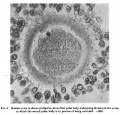 | 1,443 × 1,387 (486 KB) | Z8600021 (talk | contribs) | |
You cannot overwrite this file.
File usage
The following page uses this file:
This file contains additional information, probably added from the digital camera or scanner used to create or digitise it.
If the file has been modified from its original state, some details may not fully reflect the modified file.

