File:Stomach histology 006.jpg
From Embryology

Size of this preview: 750 × 600 pixels. Other resolution: 1,280 × 1,024 pixels.
Original file (1,280 × 1,024 pixels, file size: 227 KB, MIME type: image/jpeg)
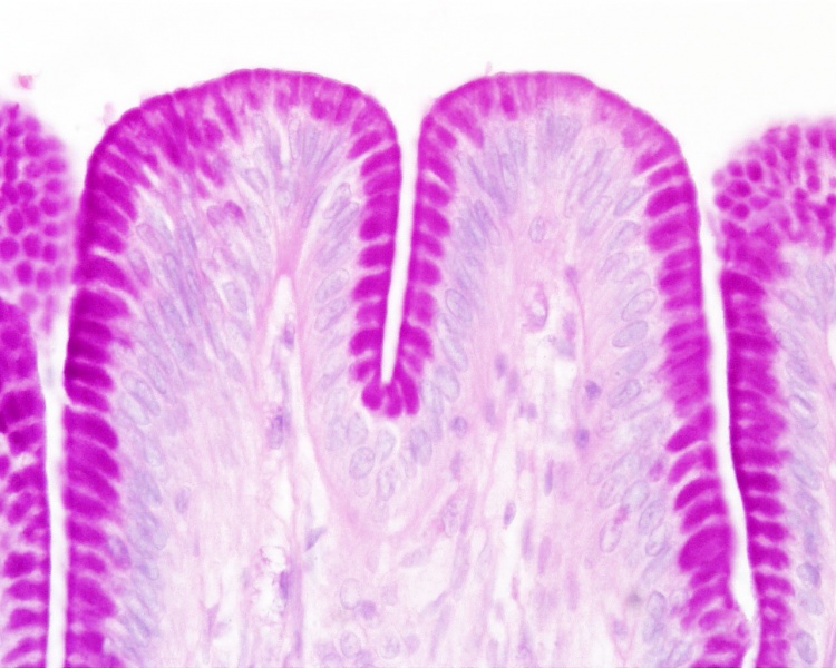
Stomach Histology
(Stain - Periodic acid-Schiff)(Stain - Haematoxylin Eosin)
- Cat stomach mucosa, secretory epithelial sheath, goblet cell, simple columnar epithelium.
- Stomach Histology Links: stomach labeled overview | parietal cells - chief cells | mucus neck - parietal cells - chief cells | stomach overview | stomach mucosa | mucosa - secretory epithelial sheath - goblet cell | gastric glands - parietal cells - chief cells | stomach overview | Stomach Histology | Stomach Development | Gastrointestinal Tract Development
Links: Histology | Histology Stains | Blue Histology images copyright Lutz Slomianka 1998-2009. The literary and artistic works on the original Blue Histology website may be reproduced, adapted, published and distributed for non-commercial purposes. See also the page Histology Stains.
Cite this page: Hill, M.A. (2024, May 15) Embryology Stomach histology 006.jpg. Retrieved from https://embryology.med.unsw.edu.au/embryology/index.php/File:Stomach_histology_006.jpg
- © Dr Mark Hill 2024, UNSW Embryology ISBN: 978 0 7334 2609 4 - UNSW CRICOS Provider Code No. 00098G
Sto40pa.jpg
File history
Click on a date/time to view the file as it appeared at that time.
| Date/Time | Thumbnail | Dimensions | User | Comment | |
|---|---|---|---|---|---|
| current | 11:58, 5 April 2013 | | 1,280 × 1,024 (227 KB) | Z8600021 (talk | contribs) | ==Stomach Histology== {{HE}} {{Stomach Histology}} {{Blue Histology}} Sto40pa.jpg |
You cannot overwrite this file.