File:Stage 19 ear.jpg

Original file (1,200 × 786 pixels, file size: 116 KB, MIME type: image/jpeg)
Human Embryo Week 7 Carnegie stage 17
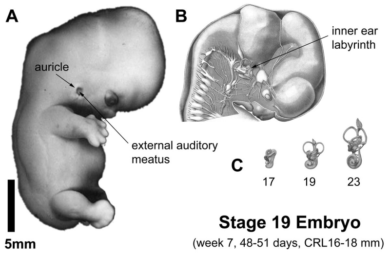
Stage 19 embryo (week 7) showing the ear development features.
A. Lateral view of the whole embryo with 5-mm scale bar. Note the pharyngeal arches have differentiated and are no longer visible on the surface. The external ear (auricle) has formed from hillocks on pharyngeal arch one and arch two. Note the relative position of the ear just above the neck and at the level of the lower jaw. The external auditory meatus is enlarged and ends at a meatal plug.
B. Historic image (Thyng, 1914) cutaway view of same stage embryo showing the position and appearance of the inner ear relative to the developing brain and other cranial ganglia.
C. The relative size and shape of the inner ear labyrinth at weeks 6, 7, and 8; by the end of the embryonic period (week 8) it approximates the shape of the adult structure.
File history
Click on a date/time to view the file as it appeared at that time.
| Date/Time | Thumbnail | Dimensions | User | Comment | |
|---|---|---|---|---|---|
| current | 18:39, 6 May 2011 | | 1,200 × 786 (116 KB) | S8600021 (talk | contribs) | ==Human Embryo Week 7 Carnegie stage 17== |
You cannot overwrite this file.
File usage
The following page uses this file: