File:Stage14 somites limbbuds.png
From Embryology
No higher resolution available.
Stage14_somites_limbbuds.png (300 × 245 pixels, file size: 24 KB, MIME type: image/png)
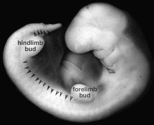
Human Embryo Carnegie Stage 14
Facts: Week 5, 31 - 35 days, 5 - 7 mm
View: Lateral left view. Amniotic membrane removed.
- Somite location shown by arrows.
- Upper and lower limb buds are labeled.
- Links: Carnegie stage 14 | Limb Development
- Carnegie Stages: 1 | 2 | 3 | 4 | 5 | 6 | 7 | 8 | 9 | 10 | 11 | 12 | 13 | 14 | 15 | 16 | 17 | 18 | 19 | 20 | 21 | 22 | 23 | About Stages | Timeline
Cite this page: Hill, M.A. (2024, May 16) Embryology Stage14 somites limbbuds.png. Retrieved from https://embryology.med.unsw.edu.au/embryology/index.php/File:Stage14_somites_limbbuds.png
- © Dr Mark Hill 2024, UNSW Embryology ISBN: 978 0 7334 2609 4 - UNSW CRICOS Provider Code No. 00098G
File history
Click on a date/time to view the file as it appeared at that time.
| Date/Time | Thumbnail | Dimensions | User | Comment | |
|---|---|---|---|---|---|
| current | 10:49, 10 August 2009 | | 300 × 245 (24 KB) | MarkHill (talk | contribs) | Image source: UNSW Embryology http://embryology.med.unsw.edu.au/Notes/skmus.htm#Somite1 Category:Mesoderm Category:Somite |
You cannot overwrite this file.
File usage
The following 10 pages use this file:
- 2009 Lecture 14
- 2009 Lecture 5
- 2010 BGD Lecture - Development of the Embryo/Fetus 2
- 2010 Lecture 14
- 2010 Lecture 5
- BGDA Lecture - Development of the Embryo/Fetus 2
- Lecture - Limb Development
- Lecture - Mesoderm Development
- Musculoskeletal System - Appendicular Skeleton Development
- Musculoskeletal System - Limb Development