File:Stage10 neural sm.jpg
Stage10_neural_sm.jpg (665 × 499 pixels, file size: 22 KB, MIME type: image/jpeg)
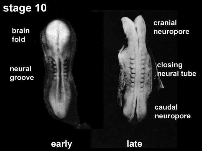
Human Embryo Week 4 - Neural Plate to Tube
Week 4 (22 - 23 days), Carnegie stage 10, 2 - 3.5 mm, Somite Number 4 - 12
View: This is a similar dorsal view of two separate embryos, amniotic membrane removed. Left embryo is an early stage 10 with broad neural plate. Right embryo is late stage 10 with plate folding to a neural groove.
Features: Somite Number 4 - 12, rostral neuropore, neural folds in region of developing brain, neural tube, somites, caudal neuropore, neural fold fuses, remnant of amniotic sac
Identify: rostral neuropore, neural folds in region of developing brain, neural tube, somites (note the different number formed), caudal neuropore, neural fold fuses, cut edge of amniotic sac
Events
Ectoderm: Neural fold deeepens, edges approach midline, neural fold fuses, neural plate folds ventrally in brain region
Mesoderm: Somitogenesis, continued segmentation of paraxial mesoderm (4 - 12 somite pairs)
| Stage 10 Links: Week 4 | Gastrulation | Lecture | Practical | Image Gallery | Carnegie Embryos | Embryos | Category:Carnegie Stage 10 | Next Stage 11 |
| Historic Papers: 1910 | 1917 | 1926 | 1939 | 1943 | 1957 | 1985 |
| Week: | 1 | 2 | 3 | 4 | 5 | 6 | 7 | 8 |
| Carnegie stage: | 1 2 3 4 | 5 6 | 7 8 9 | 10 11 12 13 | 14 15 | 16 17 | 18 19 | 20 21 22 23 |
Cite this page: Hill, M.A. (2024, May 16) Embryology Stage10 neural sm.jpg. Retrieved from https://embryology.med.unsw.edu.au/embryology/index.php/File:Stage10_neural_sm.jpg
- © Dr Mark Hill 2024, UNSW Embryology ISBN: 978 0 7334 2609 4 - UNSW CRICOS Provider Code No. 00098G
File history
Click on a date/time to view the file as it appeared at that time.
| Date/Time | Thumbnail | Dimensions | User | Comment | |
|---|---|---|---|---|---|
| current | 14:28, 10 August 2009 | | 665 × 499 (22 KB) | MarkHill (talk | contribs) | Carnegie stage 10 small image showing neuralation About Carnegie stage 10 Facts: Week 4, 22 - 23 days, 2 - 3.5 mm, Somite Number 4 - 12 View: This is a dorsal view of the embryo. Top embryo is an early stage 10, bottom is late stage 10. Amniotic membra |
You cannot overwrite this file.
File usage
The following 17 pages use this file:
- 2009 Lecture 6
- 2010 BGD Lecture - Development of the Embryo/Fetus 2
- 2010 Lecture 6
- Abnormal Development - Folic Acid and Neural Tube Defects
- BGDA Lecture - Development of the Embryo/Fetus 2
- BGDA Lecture - Development of the Nervous System
- Ectoderm
- Human System Development
- Lecture - Ectoderm Development
- Neural - Amygdala Development
- Neural - Basal Ganglia Development
- Neural - Cerebellum Development
- Neural - Spinal Cord Development
- Neural - Tectum Development
- Neural System - Abnormalities
- Neural System Development
- Talk:BGDA Lecture - Development of the Nervous System