File:Rugh 063.jpg

Original file (1,200 × 755 pixels, file size: 173 KB, MIME type: image/jpeg)
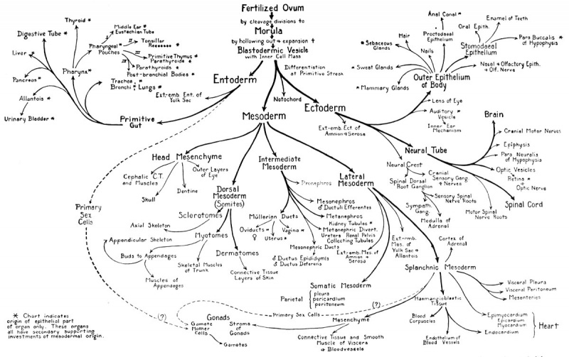
Derivatives of the Primary Germ Layers
Gastrulation is a critical stage in the development of any embryo. This is due in part to the fact that the positional relationship of the various cells of the late blastula and early gastrula begins to take on special significance. This has been demonstrated by many experimental embryologists, among whom are His, Born, BUtschli, Rhumbler, Spek, Vogt, Dalcq, Pasteels, Vintemberger, Holtfreter, Nicholas, and Schechtman. They have used various experimental devices, such as injury or excision of cell areas, or staining local areas with vital dyes and following their subsequent movement. By such methods the socalled "fate maps" of various blastulae have been worked out.
A fate map is simply a topographical surface mapping of the blastula with respect to the ultimate fate of the various areas. When one traces a cell group on the surface of a blastula which has been vitally stained with Nile blue sulfate, for instance, and finds that these cells move from the marginal zone (between the animal and the vegetal hemispheres) over the dorsal lip of the early blastopore and into the embryo where they become pharyngeal endoderm, he is then able to label the fate of that area on other blastulae. The fate maps of various amphibia are basically alike, but they are not sufficiently alike in detail to permit plotting a universal amphibian fate map. The frog's egg is very dark and it is difficult to apply vital dyes so that they will be visible on the frog blastula. However, the fate maps of closely related species have been worked out and, combined with circumstantial experimental evidence on the frog's egg, we are able to supply what is believed to be a reasonably accurate fate map of the frog blastula.
Reference
Courtesy, Patten: "Embryology of the Pig", Philadelphia, The Blakiston Company. 1927
| Historic Disclaimer - information about historic embryology pages |
|---|
 |
Reference
Rugh R. Book - The Frog Its Reproduction and Development. (1951) The Blakiston Company.
Cite this page: Hill, M.A. (2024, May 16) Embryology Rugh 063.jpg. Retrieved from https://embryology.med.unsw.edu.au/embryology/index.php/File:Rugh_063.jpg
- © Dr Mark Hill 2024, UNSW Embryology ISBN: 978 0 7334 2609 4 - UNSW CRICOS Provider Code No. 00098G
File history
Click on a date/time to view the file as it appeared at that time.
| Date/Time | Thumbnail | Dimensions | User | Comment | |
|---|---|---|---|---|---|
| current | 12:02, 12 April 2013 | | 1,200 × 755 (173 KB) | Z8600021 (talk | contribs) | {{Rugh1951 footer}} |
You cannot overwrite this file.
File usage
The following 2 pages use this file: