File:Reconstructed rat embryos 05.png
Reconstructed_rat_embryos_05.png (538 × 600 pixels, file size: 680 KB, MIME type: image/png)
Rat Blastocyst
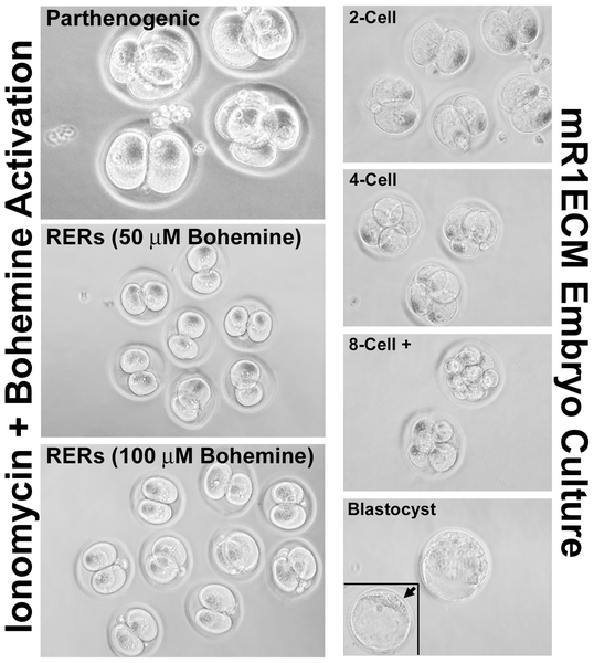
Left panels illustrate parthenogenic and reconstructed embryo activation using the calcium ionophore ionomycin followed by bohemine treatment.
There were no apparent differences between oocytes treated with 50 µM versus 100 µM bohemine, and activated parthenotes were indistinguishable from activated reconstructed rat embryos (RERs).
Right panels illustrate the development of rat embryos in mR1ECM medium. Less than 5% of rat embryos develop beyond the 4-cell stage, with only ~1% developing to the blastocyst stage (bottom panel, insert: arrow indicates blastocyst inner cell mass).
Journal.pone.0009799.g005.png
Reference
<pubmed>20333307 </pubmed>| PLoS One.
Citation: Webb RL, Findlay KA, Green MA, Beckett TL, Murphy MP (2010) Efficient Activation of Reconstructed Rat Embryos by Cyclin-Dependent Kinase Inhibitors. PLoS ONE 5(3): e9799. doi:10.1371/journal.pone.0009799
Copyright: © 2010 Webb et al. This is an open-access article distributed under the terms of the Creative Commons Attribution License, which permits unrestricted use, distribution, and reproduction in any medium, provided the original author and source are credited.
File history
Click on a date/time to view the file as it appeared at that time.
| Date/Time | Thumbnail | Dimensions | User | Comment | |
|---|---|---|---|---|---|
| current | 07:39, 29 March 2010 | | 538 × 600 (680 KB) | S8600021 (talk | contribs) | Left panels illustrate parthenogenic and reconstructed embryo activation using the calcium ionophore ionomycin followed by bohemine treatment. There were no apparent differences between oocytes treated with 50 µM versus 100 µM bohemine, and activated |
You cannot overwrite this file.
File usage
The following page uses this file: