File:Rat neural cadherin 01.jpg

Original file (1,154 × 470 pixels, file size: 62 KB, MIME type: image/jpeg)
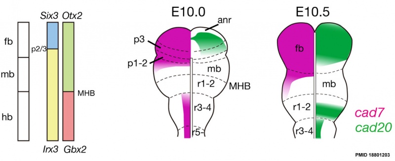
Rat Neural Cadherin Overview
Schematic illustrations of mapping of cad7 and cad20 in the early stages.
pos, pre otic sulcus; fb, forebrain; mb, midbrain; hb, hindbrain, anr, anterior neural ridge; MHB, midbrain/hindbrain boundary.
Reference
Takahashi M & Osumi N. (2008). Expression study of cadherin7 and cadherin20 in the embryonic and adult rat central nervous system. BMC Dev. Biol. , 8, 87. PMID: 18801203 DOI.
Copyright
2008 Takahashi and Osumi; licensee BioMed Central Ltd.
This is an Open Access article distributed under the terms of the Creative Commons Attribution License (http://creativecommons.org/licenses/by/2.0), which permits unrestricted use, distribution, and reproduction in any medium, provided the original work is properly cited.
1471-213X-8-87-3.jpg
Cite this page: Hill, M.A. (2024, May 8) Embryology Rat neural cadherin 01.jpg. Retrieved from https://embryology.med.unsw.edu.au/embryology/index.php/File:Rat_neural_cadherin_01.jpg
- © Dr Mark Hill 2024, UNSW Embryology ISBN: 978 0 7334 2609 4 - UNSW CRICOS Provider Code No. 00098G
File history
Click on a date/time to view the file as it appeared at that time.
| Date/Time | Thumbnail | Dimensions | User | Comment | |
|---|---|---|---|---|---|
| current | 18:09, 23 June 2012 | 1,154 × 470 (62 KB) | Z8600021 (talk | contribs) | ==Rat Neural Cadherin Overview== Schematic illustrations of mapping of cad7 and cad20 in the early stages. pos, pre otic sulcus; fb, forebrain; mb, midbrain; hb, hindbrain, anr, anterior neural ridge; MHB, midbrain/hindbrain boundary. ===Reference=== |
You cannot overwrite this file.
File usage
The following 2 pages use this file: