File:Postnatal persistant ductus venosus ultrasound 03.jpg

Postnatal_persistant_ductus_venosus_ultrasound_03.jpg (694 × 600 pixels, file size: 54 KB, MIME type: image/jpeg)
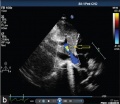
Persistent Ductus Venosus
| Persistent ductus venosus (postnatal 8 years) connecting the left portal vein to the inferior vena cava. Subcostal two-dimensional echocardiography (yellow arrow)
|
|
Reference
<pubmed>24688239</pubmed>| Ann Pediatr Cardiol.
Copyright
© 2008 Annals of Pediatric Cardiology
The entire contents of the Annals of Pediatric Cardiology are protected under Indian and international copyrights. The Journal, however, grants to all users a free, irrevocable, worldwide, perpetual right of access to, and a license to copy, use, distribute, perform and display the work publicly and to make and distribute derivative works in any digital medium for any reasonable non-commercial purpose, subject to proper attribution of authorship and ownership of the rights.
Figure 1 Original figure resized and relabelled.
File history
Click on a date/time to view the file as it appeared at that time.
| Date/Time | Thumbnail | Dimensions | User | Comment | |
|---|---|---|---|---|---|
| current | 11:28, 21 February 2015 | | 694 × 600 (54 KB) | Z8600021 (talk | contribs) |
You cannot overwrite this file.
File usage
The following 11 pages use this file:
- ANAT2341 Lab 11 - Second Trimester
- BGDA Practical 12 - Second Trimester
- Cardiovascular System - Ductus Venosus
- Movies
- Patent Ductus Venosus Movie
- Ultrasound
- Ultrasound - Abnormal
- File:Postnatal persistant ductus venosus ultrasound 03.jpg
- Template:Ultrasound Movies
- Template:Ultrasound Movies - abnormal
- Template:Ultrasound patent ductus venosus movie