File:Placental cord epithelium 01.jpg
From Embryology
Size of this preview: 800 × 537 pixels. Other resolution: 1,200 × 805 pixels.
Original file (1,200 × 805 pixels, file size: 130 KB, MIME type: image/jpeg)
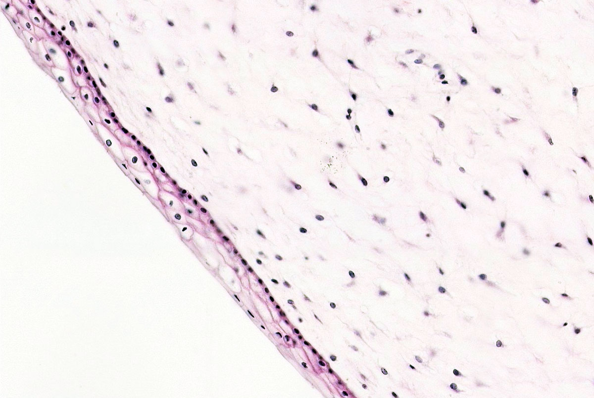
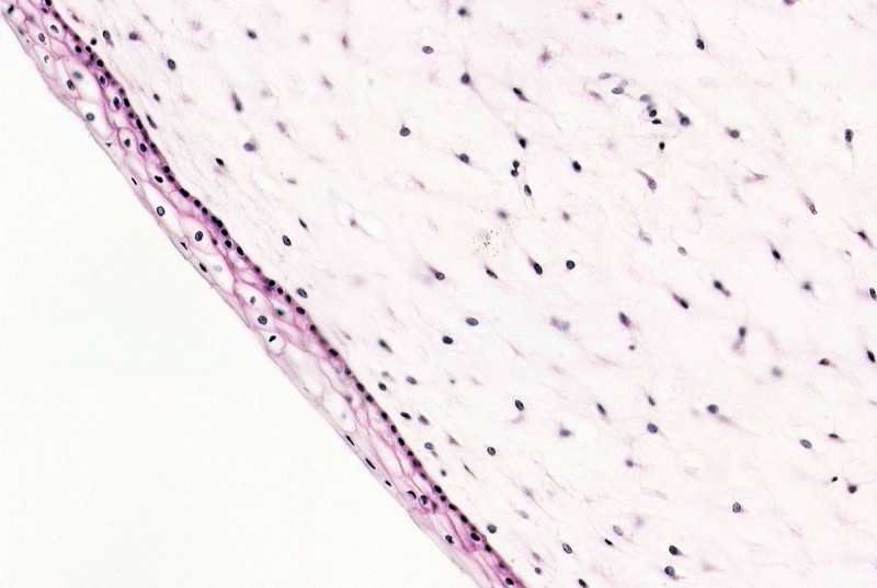
Placental Cord Histology
This histological section shows the typical epithelial (stratified squamous) structure found on the surface of the placental (umbilical) cord. Note that the appearance in this post-birth placental cord is quite different from that before birth.
- Underlying the epithelium is the expanded connective tissue of the placental cord.
- External to the epithelium would be the amniotic fluid.
- Placental Cord Histology: Cord overview | Vein | Artery | Artery | Allantois | Epithelium | Cord overview 1 unlabeled | overview 2 unlabeled | unlabeled vein and connective tissue | unlabeled connective tissue | Villi histology | Placenta Histology
Source: UNSW Embryology
File history
Click on a date/time to view the file as it appeared at that time.
| Date/Time | Thumbnail | Dimensions | User | Comment | |
|---|---|---|---|---|---|
| current | 15:33, 31 July 2011 | | 1,200 × 805 (130 KB) | S8600021 (talk | contribs) | ==Placental Cord Histology== This histological section shows the typical structures found in the placental (umbilical) cord. Note that the appearance in this post-birth placental cord is quite different from that before birth. Source: UNSW Embryology [ |
You cannot overwrite this file.
File usage
The following page uses this file: