File:Pituitary histology 011.jpg
From Embryology
Size of this preview: 389 × 600 pixels. Other resolution: 900 × 1,388 pixels.
Original file (900 × 1,388 pixels, file size: 309 KB, MIME type: image/jpeg)
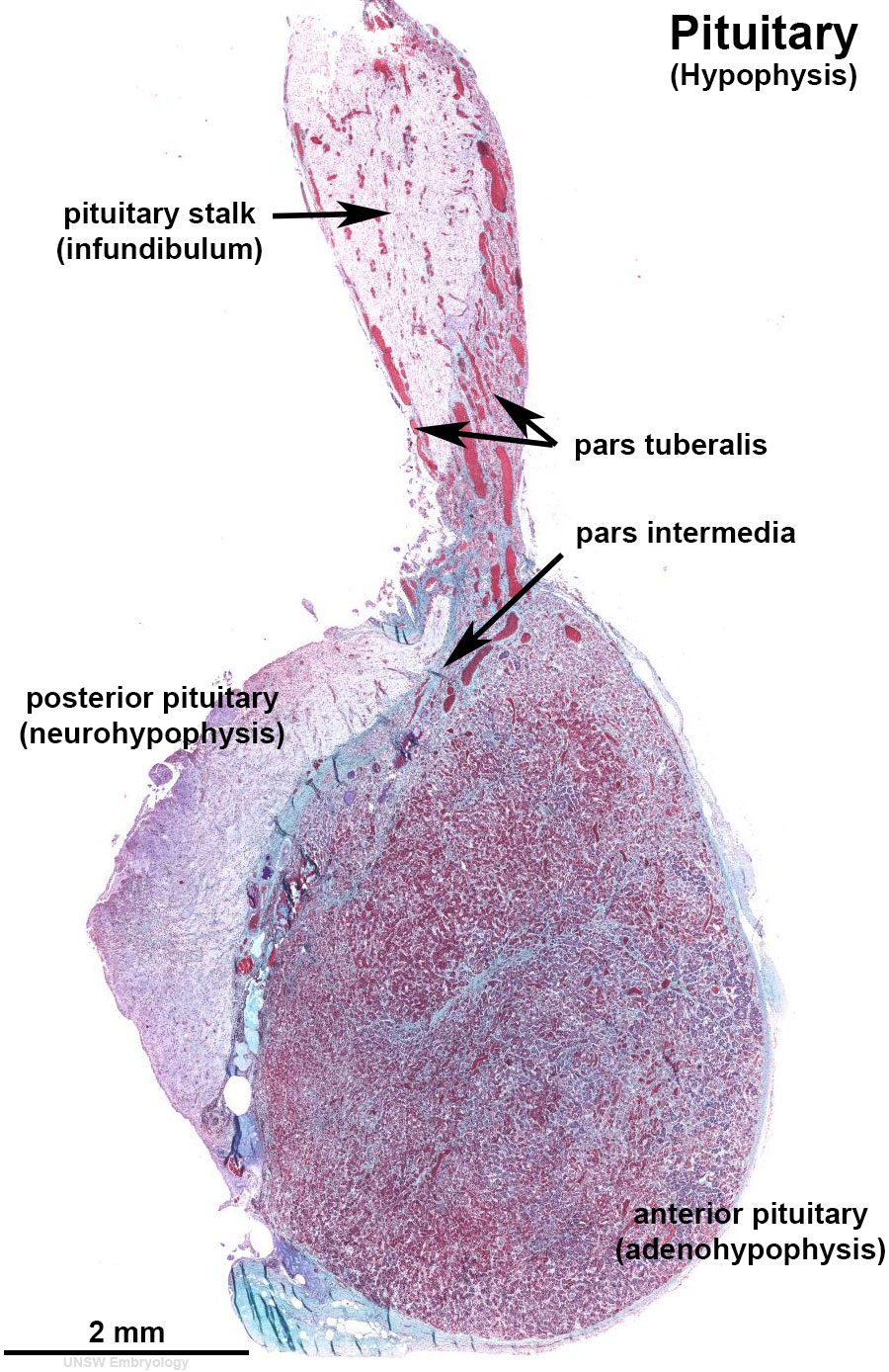
Pituitary (Hypophysis) Histology
This overview histology image shows a sagittal section through the adult pituitary (hypophysis). The major regions of this endocrine organ are identified by labels. Scale bar is 2 mm.
- Pituitary Histology: Pituitary overview | Anterior H&E | Anterior H&E | Anterior labeled | PAS/O Overview | Acidophils | Basophils | Posterior labeled | Posterior unlabeled | Histology Stains | BGD - Endocrine Histology | Pituitary Development
Cite this page: Hill, M.A. (2024, April 27) Embryology Pituitary histology 011.jpg. Retrieved from https://embryology.med.unsw.edu.au/embryology/index.php/File:Pituitary_histology_011.jpg
- © Dr Mark Hill 2024, UNSW Embryology ISBN: 978 0 7334 2609 4 - UNSW CRICOS Provider Code No. 00098G
File history
Click on a date/time to view the file as it appeared at that time.
| Date/Time | Thumbnail | Dimensions | User | Comment | |
|---|---|---|---|---|---|
| current | 10:44, 11 November 2016 | | 900 × 1,388 (309 KB) | Z8600021 (talk | contribs) | ==Pituitary (Hypophysis) Histology (adult)== {{Footer}} Category:Indexed pages Category:Human Category:Histology Category:Endocrine |
You cannot overwrite this file.
File usage
The following 3 pages use this file: