File:Mouse thymus development 03.jpg
Original file (1,200 × 487 pixels, file size: 260 KB, MIME type: image/jpeg)
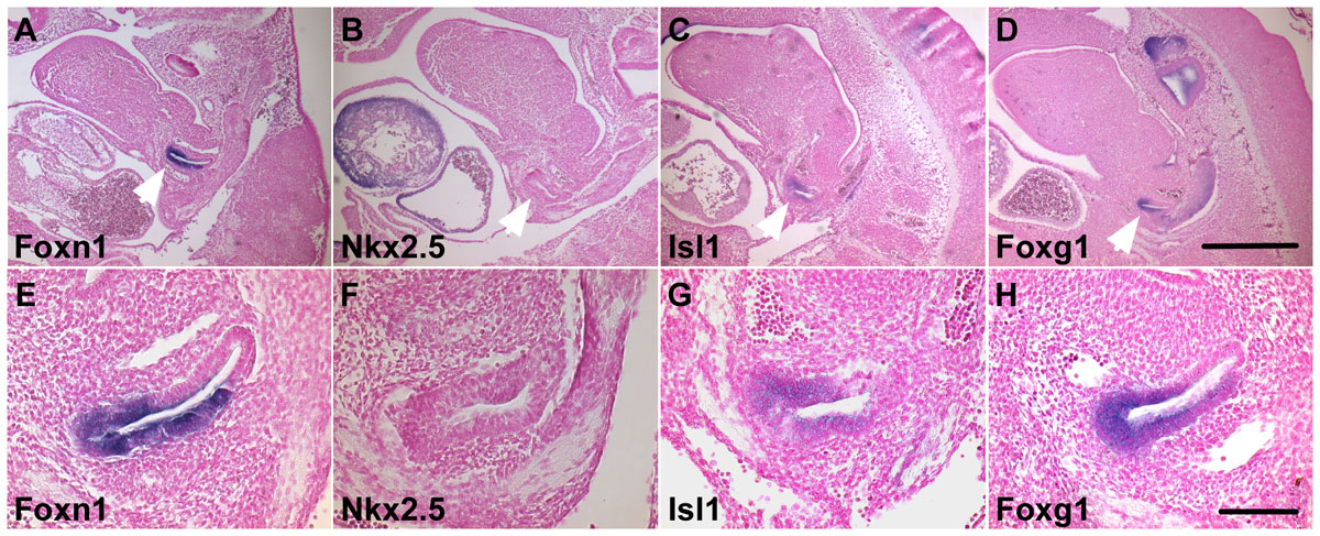
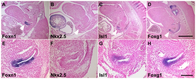
Mouse Thymus Expression of Isl1 and Foxg1 in the ventral third pouch endoderm/thymus rudiment at E11.5
Parasagittal sections of E11.5 embryos hybridized with probes for:
- Foxn1 (A, E)
- Nkx2-5 (B, F)
- Isl1 (C, G)
- Foxg1 (D, H)
Ventral is on the left and anterior is up.
Arrow heads in A–D indicate the third pouch.
Scale bar represents 500 (A–D) and 100 µm (E–H).
Reference
<pubmed>22087235</pubmed>| PLoS One.
© 2011 Wei, Condie. This is an open-access article distributed under the terms of the Creative Commons Attribution License, which permits unrestricted use, distribution, and reproduction in any medium, provided the original author and source are credited.
Original file name: Figure 4. Journal.pone.0026795.g004.png
doi:10.1371/journal.pone.0026795.g004
Mouse thymus development 01.jpg
File history
Click on a date/time to view the file as it appeared at that time.
| Date/Time | Thumbnail | Dimensions | User | Comment | |
|---|---|---|---|---|---|
| current | 13:22, 20 February 2012 | 1,200 × 487 (260 KB) | S8600021 (talk | contribs) | ===Reference=== <pubmed>22087235</pubmed>| [http://www.plosone.org/article/info%3Adoi%2F10.1371%2Fjournal.pone.0026795 PLoS One.] © 2011 Wei, Condie. This is an open-access article distributed under the terms of the Creative Commons Attribution Licens |
You cannot overwrite this file.
File usage
The following page uses this file: