File:Mouse neural tube bending model.jpg
From Embryology

Size of this preview: 800 × 377 pixels. Other resolution: 1,000 × 471 pixels.
Original file (1,000 × 471 pixels, file size: 79 KB, MIME type: image/jpeg)
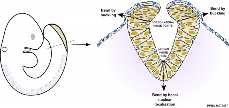
Mouse Neural Tube Bending Model
The closing mouse spinal neural plate bends at the midline and dorsolaterally.
- Basal nuclear localisation causes midline, but not dorsolateral, bending.
- Neuroepithelial cells translocate into the dorsolateral neural plate during closure.
- A discontinuity of cell density results between dorsal and ventral neural plate.
- Buckling at this discontinuity may result in dorsolateral neural plate bending.
Reference
<pubmed>26079577</pubmed>| PMC4528075 | Dev Biol.
Dev Biol. 2015 Aug 15; 404(2): 113–124. doi: 10.1016/j.ydbio.2015.06.003
Copyright
http://creativecommons.org/licenses/by/4.0
Image resized and PMID label added.
Cite this page: Hill, M.A. (2024, May 4) Embryology Mouse neural tube bending model.jpg. Retrieved from https://embryology.med.unsw.edu.au/embryology/index.php/File:Mouse_neural_tube_bending_model.jpg
- © Dr Mark Hill 2024, UNSW Embryology ISBN: 978 0 7334 2609 4 - UNSW CRICOS Provider Code No. 00098G
File history
Click on a date/time to view the file as it appeared at that time.
| Date/Time | Thumbnail | Dimensions | User | Comment | |
|---|---|---|---|---|---|
| current | 15:36, 16 August 2015 | | 1,000 × 471 (79 KB) | Z8600021 (talk | contribs) | ===Cellular basis of neuroepithelial bending during mouse spinal neural tube closure=== Dev Biol. 2015 Aug 15;404(2):113-24. doi: 10.1016/j.ydbio.2015.06.003. Epub 2015 Jun 12. McShane SG1, Molè MA1, Savery D1, Greene ND1, Tam PP2, Copp AJ3. Abstra... |
You cannot overwrite this file.
File usage
The following page uses this file: