File:Liver histology EM02.jpg

Original file (1,028 × 707 pixels, file size: 154 KB, MIME type: image/jpeg)
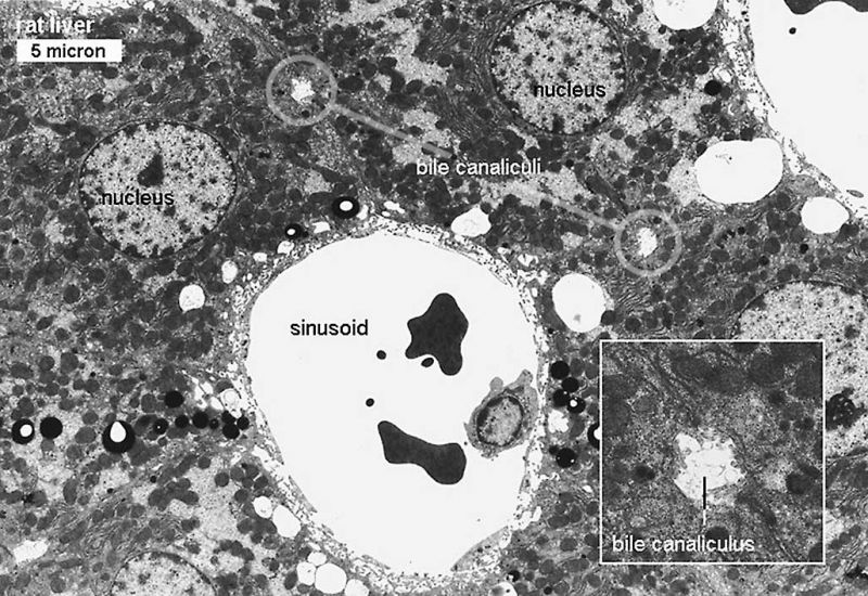
Rat Liver Histology (Electron Micrograph)
This electron micrograph shows the relative size and position of the liver sinusoidal space and the small canalicul running between the hepatocytes.
- Links: Liver histology EM01 | Liver histology EM02 | Liver Histology | liver
Links: Histology | Histology Stains | Blue Histology images copyright Lutz Slomianka 1998-2009. The literary and artistic works on the original Blue Histology website may be reproduced, adapted, published and distributed for non-commercial purposes. See also the page Histology Stains.
Cite this page: Hill, M.A. (2024, May 21) Embryology Liver histology EM02.jpg. Retrieved from https://embryology.med.unsw.edu.au/embryology/index.php/File:Liver_histology_EM02.jpg
- © Dr Mark Hill 2024, UNSW Embryology ISBN: 978 0 7334 2609 4 - UNSW CRICOS Provider Code No. 00098G
File history
Click on a date/time to view the file as it appeared at that time.
| Date/Time | Thumbnail | Dimensions | User | Comment | |
|---|---|---|---|---|---|
| current | 14:19, 24 July 2019 | | 1,028 × 707 (154 KB) | Z8600021 (talk | contribs) | Reverted to version as of 20:47, 26 April 2018 (AEST) |
| 14:18, 24 July 2019 |  | 1,745 × 1,200 (469 KB) | Z8600021 (talk | contribs) | ||
| 20:47, 26 April 2018 | 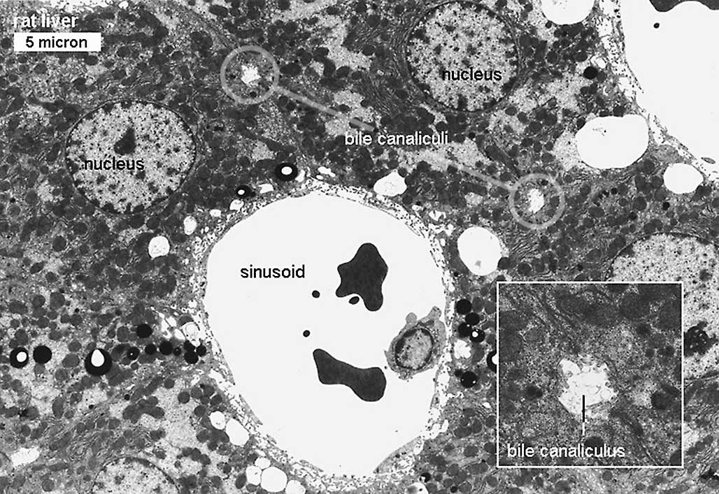 | 1,028 × 707 (154 KB) | Z8600021 (talk | contribs) | ||
| 16:50, 11 August 2011 |  | 800 × 551 (159 KB) | S8600021 (talk | contribs) |
You cannot overwrite this file.
File usage
The following 2 pages use this file: