File:Licata1954 fig04.jpg
From Embryology

Size of this preview: 784 × 600 pixels. Other resolution: 1,000 × 765 pixels.
Original file (1,000 × 765 pixels, file size: 84 KB, MIME type: image/jpeg)
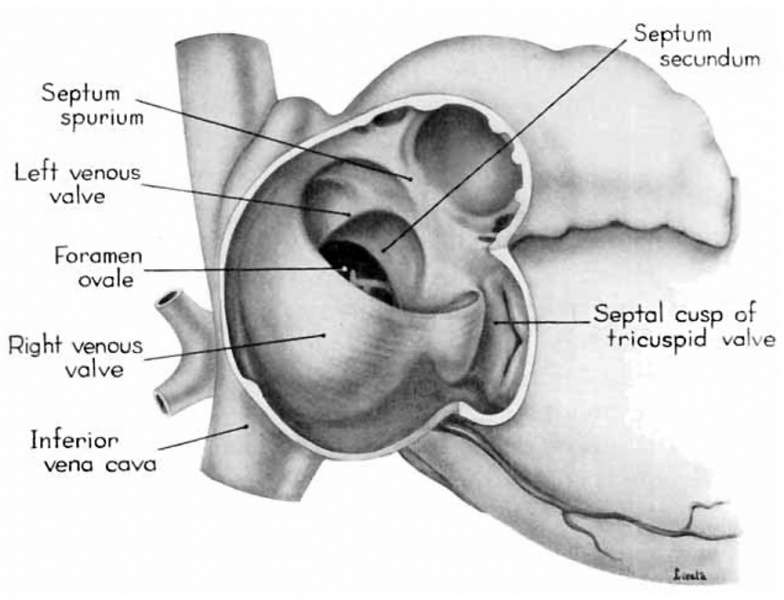
Fig. 4. Dextral View of heart of 25 mm embryo with atrium opened
A tag of tissue which is attached to the septum primum is just visible above the right venous valve.
(Reconstruction X 100, illustration X 40.)
- Links: fig 1 | fig 2 | fig 3 | fig 4 | fig 5 | fig 6 | fig 7 | fig 8 | fig 9 | fig 10 | fig 11 | fig 12 | fig 13 | fig 14 | fig 15 | fig 16 | fig 16a | fig 16b | fig 16c | fig 16d | 1954 Licata | Historic Papers | Heart Development
Reference
Licata RH. The human embryonic heart in the ninth week. (1954) Amer. J Anat., 94: 73-125. PMID 13124266
Cite this page: Hill, M.A. (2024, May 9) Embryology Licata1954 fig04.jpg. Retrieved from https://embryology.med.unsw.edu.au/embryology/index.php/File:Licata1954_fig04.jpg
- © Dr Mark Hill 2024, UNSW Embryology ISBN: 978 0 7334 2609 4 - UNSW CRICOS Provider Code No. 00098G
File history
Click on a date/time to view the file as it appeared at that time.
| Date/Time | Thumbnail | Dimensions | User | Comment | |
|---|---|---|---|---|---|
| current | 10:48, 5 March 2017 | | 1,000 × 765 (84 KB) | Z8600021 (talk | contribs) | |
| 10:47, 5 March 2017 | 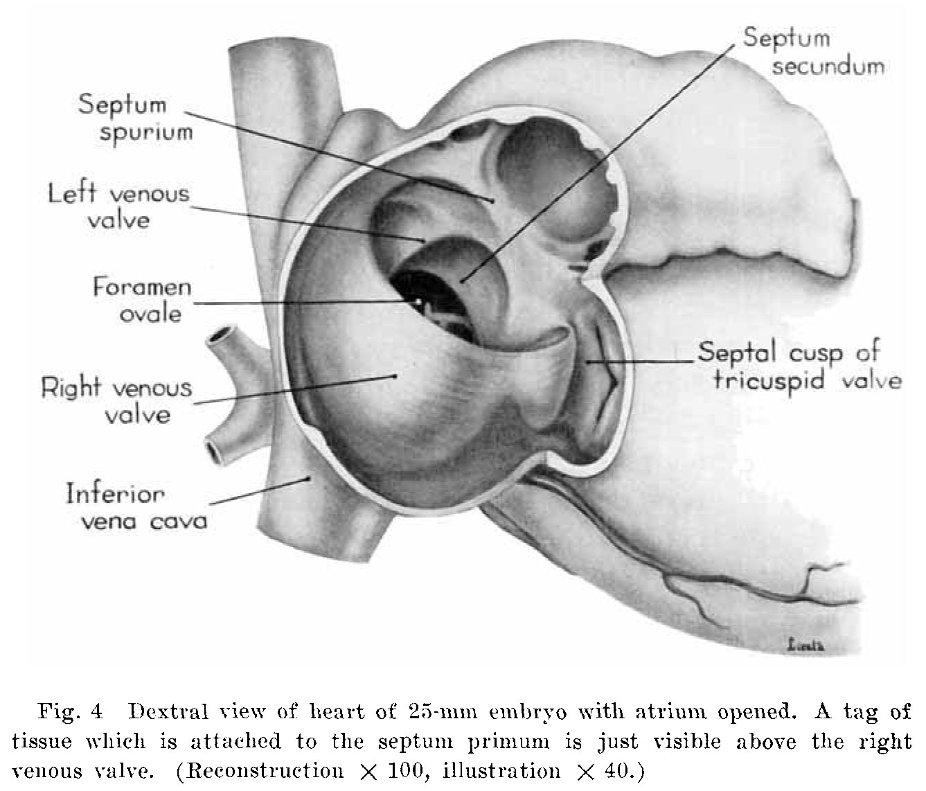 | 1,337 × 1,142 (174 KB) | Z8600021 (talk | contribs) |
You cannot overwrite this file.
File usage
The following 4 pages use this file: