File:Kollmann537.jpg
Kollmann537.jpg (481 × 489 pixels, file size: 45 KB, MIME type: image/jpeg)
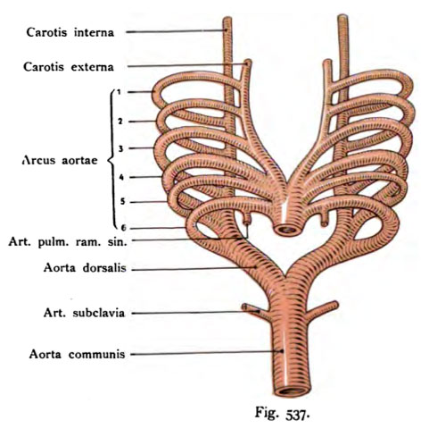
Fig. 537. Aortic arch of mammals and man
shown schematically.
The origin of the truncus arteriosus, the curve of the aortic arch, continued in the aortic roots and the origin of the dorsal aorta. Cf. Fig. 535 of a cartilaginous fish.
There are six in reptiles, ranging from the aortic arch on each side in all mammals studied so far been demonstrated, even in man.
- This text is a Google translate computer generated translation and may contain many errors.
Images from - Atlas of the Development of Man (Volume 2)
(Handatlas der entwicklungsgeschichte des menschen)
- Kollmann Atlas 2: Gastrointestinal | Respiratory | Urogenital | Cardiovascular | Neural | Integumentary | Smell | Vision | Hearing | Kollmann Atlas 1 | Kollmann Atlas 2 | Julius Kollmann
- Links: Julius Kollman | Atlas Vol.1 | Atlas Vol.2 | Embryology History
| Historic Disclaimer - information about historic embryology pages |
|---|
 |
Reference
Kollmann JKE. Atlas of the Development of Man (Handatlas der entwicklungsgeschichte des menschen). (1907) Vol.1 and Vol. 2. Jena, Gustav Fischer. (1898).
Cite this page: Hill, M.A. (2024, May 9) Embryology Kollmann537.jpg. Retrieved from https://embryology.med.unsw.edu.au/embryology/index.php/File:Kollmann537.jpg
- © Dr Mark Hill 2024, UNSW Embryology ISBN: 978 0 7334 2609 4 - UNSW CRICOS Provider Code No. 00098G
Fig. 537. Aortenbogen der Säuger und des Menschen
schematisch dargestellt.
Der Ursprung aus dem Truncus arteriosus, der Verlauf der Aortenbogen, ihre Fortsetzung in die Aortenwurzeln und die Entstehung der dorsalen Aorta. Vergl. die Fig. 535 von einem Knorpelfisch.
Es sind von den Reptilien angefangen sechs Aortenbogen auf jeder Seite bei allen bisher untersuchten Säugern nachgewiesen worden. Auch bei dem Menschen.
File history
Click on a date/time to view the file as it appeared at that time.
| Date/Time | Thumbnail | Dimensions | User | Comment | |
|---|---|---|---|---|---|
| current | 00:10, 17 October 2011 | | 481 × 489 (45 KB) | S8600021 (talk | contribs) | {{Kollmann1907}} |
You cannot overwrite this file.
File usage
The following 2 pages use this file: