File:Keibel1910 fig22.jpg
Keibel1910_fig22.jpg (520 × 520 pixels, file size: 25 KB, MIME type: image/jpeg)
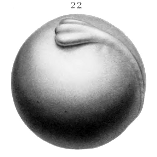
Fig. 22. Dorso-lateral view of egg 20 days 10 hrs old
Dorso-lateral view of embryo 20 days 10 hrs. old, length 6 mm, 6 pairs of myotomes. Outline of body conforms to curvature of egg. Head end of embryo shows three longitudinal ridges; middle ridge lies slightly above level of lateral ridges. The middle one is common anläge of fore, mid and bind brain. The lateral ones are the common anläge of the optic vesicles and branchial arches. Anus formed. (X 10.)
| Historic Disclaimer - information about historic embryology pages |
|---|
 |
Reference
Eycleshymer AC. and Wilson JM. Normal Plates of the Development of the Salamander Embryo (Nectürüs maculosus). Vol. 11 in series by Keibel F. Normal plates of the development of vertebrates (Normentafeln zur Entwicklungsgeschichte der Wirbelthiere) Fisher, Jena., Germany.
Cite this page: Hill, M.A. (2024, May 15) Embryology Keibel1910 fig22.jpg. Retrieved from https://embryology.med.unsw.edu.au/embryology/index.php/File:Keibel1910_fig22.jpg
- © Dr Mark Hill 2024, UNSW Embryology ISBN: 978 0 7334 2609 4 - UNSW CRICOS Provider Code No. 00098G
File history
Click on a date/time to view the file as it appeared at that time.
| Date/Time | Thumbnail | Dimensions | User | Comment | |
|---|---|---|---|---|---|
| current | 14:09, 10 January 2015 | | 520 × 520 (25 KB) | Z8600021 (talk | contribs) | ==Fig. 22. Dorso-lateral view of egg 20 days 10 hrs old== Dorso-lateral view of embryo 20 days 10 hrs. old, length 6 mm, 6 pairs of myotomes. Outline of body conforms to curvature of egg. Head end of embryo shows three longitudinal ridges; middle ridg... |
You cannot overwrite this file.
File usage
The following 3 pages use this file: