File:Inner ear haircells.jpg
Original file (800 × 671 pixels, file size: 121 KB, MIME type: image/jpeg)
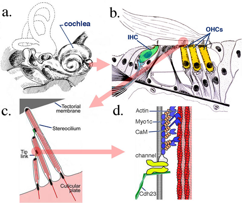
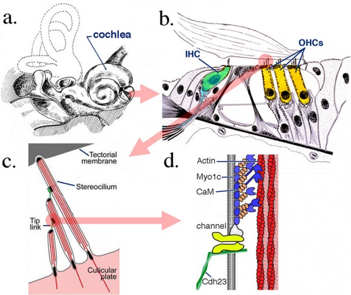
Anatomical details of inner ear
The cochlea and organ of Corti, the sense organ of mammalian hearing.
(a) The cochlea, a fluid-filled tripartite channel, is located in the inner ear
(b) A hemisected cochlea provides a radial view of the organ of Corti, a cellular matrix showing the location of hair cells. The input organelles of hair cells, the stereocilia, are connected by different links, including tip-link proteins allowing movement as a unit.
(c) Deflection of the stereocilary bundle due to displacement between the top of the organ of Corti and the bottom of the tectorial membrane provides tension to the tip link, which, in turn, modulates the cation-selective mechanoelectrical transduction (MET) channel's open probability.
(d) The tip link is partially composed of cdh23, which is presumed to interact with the MET channel either directly or indirectly.
Legend
- IHC - inner hair cell
- OHC - outer hair cell
- Myo1c - myosin 1c
- CaM - calmodulin
- Links: inner ear
Reference
Zheng J, Anderson CT, Miller KK, Cheatham M & Dallos P. (2009). Identifying components of the hair-cell interactome involved in cochlear amplification. BMC Genomics , 10, 127. PMID: 19320974 DOI.
Copyright
© 2009 Zheng et al; licensee BioMed Central Ltd.
This is an Open Access article distributed under the terms of the Creative Commons Attribution License (http://creativecommons.org/licenses/by/2.0), which permits unrestricted use, distribution, and reproduction in any medium, provided the original work is properly cited.
Original image name: 1471-2164-10-127-1-l.jpg Images in (c) and (d) are modified from LeMasurier and Gillespie PMID 16269359.
Cite this page: Hill, M.A. (2024, May 4) Embryology Inner ear haircells.jpg. Retrieved from https://embryology.med.unsw.edu.au/embryology/index.php/File:Inner_ear_haircells.jpg
- © Dr Mark Hill 2024, UNSW Embryology ISBN: 978 0 7334 2609 4 - UNSW CRICOS Provider Code No. 00098G
File history
Click on a date/time to view the file as it appeared at that time.
| Date/Time | Thumbnail | Dimensions | User | Comment | |
|---|---|---|---|---|---|
| current | 12:56, 5 October 2011 | | 800 × 671 (121 KB) | S8600021 (talk | contribs) | |
| 00:59, 28 September 2009 |  | 600 × 510 (45 KB) | S8600021 (talk | contribs) | Anatomical details of inner ear, cochlea and organ of Corti, the sense organ of mammalian hearing. The cochlea, a fluid-filled tripartite channel, is located in the inner ear (a). A hemisected cochlea provides a radial view of the organ of Corti, a cell |
You cannot overwrite this file.
File usage
The following 6 pages use this file: