File:Human pronuclear stage EM17.jpg
Original file (1,104 × 504 pixels, file size: 156 KB, MIME type: image/jpeg)
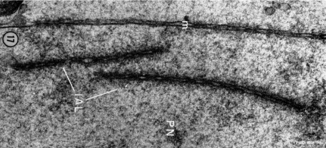
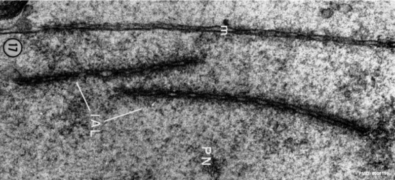
Human Pronuclear Stage - Pronuclei Annulate Lamellae
- The annulate lamellae (IAL) which are present inside the pronuelei (PN) are morphologically identical to those observed in the ooplasm (compare with Fig. 9).
- Note also the similarity between the annulate lamellae and the pronuclear envelope (m).
X 6~,000.
- Pronuclear Stage EM: | Human pronuclei | Human pronuclei | Mitochondria | Mitochondria | Endoplasmic Reticulum | Endoplasmic Reticulum | Endoplasmic Reticulum | Cytoplasmic LameIlae | Cytoplasmic LameIlae | Crystalline inclusions | Crystalline inclusion | Pronuclear Envelope | Pronuclei Annulate Lamellae | Pronucleus | Nucleoli | Spermatozoon | Human pronuclei | Spermatozoon Components | Golgi complex | First Polar Body | First Polar Body | First Polar Body | Second Polar Body | Second Polar Body | pronuclei | polar body | fertilization | Carnegie stage 1
Reference
Zamboni L, Mishell DR, Bell JH & Baca M. (1966). Fine structure of the human ovum in the pronuclear stage. J. Cell Biol. , 30, 579-600. PMID: 6008199
Copyright
Rockefeller University Press - Copyright Policy This article is distributed under the terms of an Attribution–Noncommercial–Share Alike–No Mirror Sites license for the first six months after the publication date (see http://www.jcb.org/misc/terms.shtml). After six months it is available under a Creative Commons License (Attribution–Noncommercial–Share Alike 4.0 Unported license, as described at https://creativecommons.org/licenses/by-nc-sa/4.0/ ). (More? Help:Copyright Tutorial)
Image has been scaled, rotated and contrast adjusted.
File history
Click on a date/time to view the file as it appeared at that time.
| Date/Time | Thumbnail | Dimensions | User | Comment | |
|---|---|---|---|---|---|
| current | 23:47, 23 February 2012 | | 1,104 × 504 (156 KB) | Z8600021 (talk | contribs) |
You cannot overwrite this file.
File usage
The following page uses this file: