File:Human heart SEM1.jpg
From Embryology
Size of this preview: 800 × 220 pixels. Other resolution: 1,200 × 330 pixels.
Original file (1,200 × 330 pixels, file size: 47 KB, MIME type: image/jpeg)
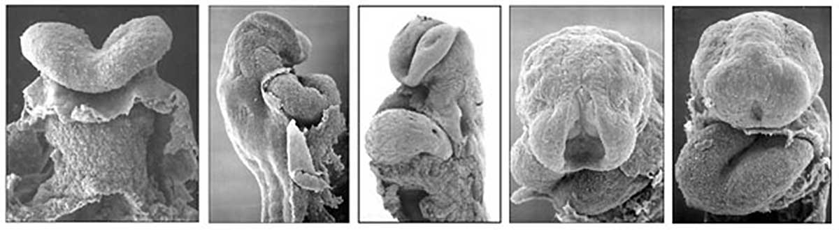
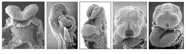
The Human Heart from day 21 to 25
Scanning electron micrograph images of the early human embryonic heart tube. Note the anterior body wall has been removed exposing the pericardial cavity in which the heart tube lies.
- Heart Links: Image day 21 to 25 | Image day 21 | Image day 25 | Carnegie stage 10 | Carnegie stage 11 | Cardiovascular System Development
Image Source: Scanning electron micrographs of the Carnegie stages of the early human embryos are reproduced with the permission of Prof Kathy Sulik, from embryos collected by Dr. Vekemans and Tania Attié-Bitach. Images are for educational purposes only and cannot be reproduced electronically or in writing without permission.
File history
Click on a date/time to view the file as it appeared at that time.
| Date/Time | Thumbnail | Dimensions | User | Comment | |
|---|---|---|---|---|---|
| current | 13:50, 24 August 2014 | 1,200 × 330 (47 KB) | Z8600021 (talk | contribs) | ||
| 21:21, 16 August 2009 | 600 × 177 (30 KB) | S8600021 (talk | contribs) | The Human Heart from day 10 to 25 |
You cannot overwrite this file.
File usage
The following 13 pages use this file:
- 2009 Lecture 7
- 2010 BGD Lecture - Development of the Embryo/Fetus 1
- 2010 Lecture 7
- ANAT2341 Lab 4 - Early Cardiovascular Development
- BGDA Lecture - Development of the Embryo/Fetus 1
- Cardiovascular System - Circulation Development
- Cardiovascular System - Coronary Circulation Development
- Cardiovascular System Development
- Fetal ECHO Meeting 2012
- Human Embryo SEM
- Lecture - Early Vascular Development
- RPAH Cardiac Embryology 2014
- User:Z3267024