File:Human Carnegie stage 1-23.jpg

Original file (1,000 × 563 pixels, file size: 98 KB, MIME type: image/jpeg)
Human Embryonic Development
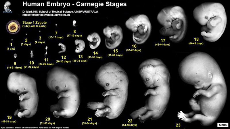
| Carnegie stage 1 to 23, showing human external appearance and growth during the first 8 weeks of development.
Stage 1 is early zygote and not in scale. Has been enlarged to show pronuclei and polar bodies. (5mm scale bar, lower right of image)
|
|
| Week: | 1 | 2 | 3 | 4 | 5 | 6 | 7 | 8 |
| Carnegie stage: | 1 2 3 4 | 5 6 | 7 8 9 | 10 11 12 13 | 14 15 | 16 17 | 18 19 | 20 21 22 23 |
- Carnegie Stages: 1 | 2 | 3 | 4 | 5 | 6 | 7 | 8 | 9 | 10 | 11 | 12 | 13 | 14 | 15 | 16 | 17 | 18 | 19 | 20 | 21 | 22 | 23 | About Stages | Timeline
Image source: The Kyoto Collection images are reproduced with the permission of Prof. Kohei Shiota and Prof. Shigehito Yamada, Anatomy and Developmental Biology, Kyoto University Graduate School of Medicine, Kyoto, Japan for educational purposes only and cannot be reproduced electronically or in writing without permission.
Smaller version of stage poster showing embryos in scale with each other.
Glossary Links
- Glossary: A | B | C | D | E | F | G | H | I | J | K | L | M | N | O | P | Q | R | S | T | U | V | W | X | Y | Z | Numbers | Symbols | Term Link
Cite this page: Hill, M.A. (2024, May 15) Embryology Human Carnegie stage 1-23.jpg. Retrieved from https://embryology.med.unsw.edu.au/embryology/index.php/File:Human_Carnegie_stage_1-23.jpg
- © Dr Mark Hill 2024, UNSW Embryology ISBN: 978 0 7334 2609 4 - UNSW CRICOS Provider Code No. 00098G
File history
Click on a date/time to view the file as it appeared at that time.
| Date/Time | Thumbnail | Dimensions | User | Comment | |
|---|---|---|---|---|---|
| current | 11:13, 14 July 2016 | | 1,000 × 563 (98 KB) | Z8600021 (talk | contribs) | |
| 14:23, 11 August 2009 | 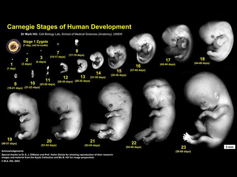 | 800 × 600 (51 KB) | MarkHill (talk | contribs) | Human embryo images Carnegie stage 1 to 23. Smaller version of stage poster showing embryos in scale with each other. Stage 1 is early zygote and not in scale. Has been enlarged to show pronuclei and polar bodies. (5mm scale bar, lower right of image) |
You cannot overwrite this file.
File usage
The following 37 pages use this file:
- 2010 BGD Lecture - Development of the Embryo/Fetus 2
- 2010 BGD Tutorial - Applied Embryology and Teratology
- 2010 Foundations Lecture - Introduction to Human Development
- 2010 Lab 6
- ANAT2341 Embryology 2010
- ANAT2341 Embryology 2011
- ANAT2341 Embryology 2012
- ANAT2341 Embryology 2013
- ANAT2341 Embryology 2014
- ANZACA Meeting 2012 - Embryology
- Anatomy and Embryology Goettingen - 2013 Seminar
- BGDA Lecture - Development of the Embryo/Fetus 2
- BGD Tutorial - Applied Embryology and Teratology
- Development Group Meeting 2017 - Digital Embryology Consortium
- Embryonic Development
- Fetal ECHO Meeting 2012
- Foundations Lecture - Introduction to Human Development
- Foundations Practical - Week 1 to 8
- Gottingen Meeting 2017 - Digital Embryology Consortium
- Human Embryo Collections
- Kyoto Collection
- Lecture - 2011 Course Introduction
- Lecture - 2012 Course Introduction
- Lecture - 2013 Course Introduction
- Lecture - 2014 Course Introduction
- Lecture - 2015 Course Introduction
- Lecture - 2016 Course Introduction
- Lecture - 2017 Course Introduction
- Lecture - Week 3 Development
- Museum of Natural History Berlin - 2013 Seminar
- Pre-Medicine Program - Embryology
- RPAH Cardiac Embryology 2014
- UNSW Learning and Teaching Seminar 2012
- Talk:Berlin Meeting 2017 - Digital Embryology Consortium
- Talk:Development Group Meeting 2017 - Digital Embryology Consortium
- Talk:Lecture - 2016 Course Introduction
- Talk:Main Page