File:Huber1915 1fig09.jpg
Huber1915_1fig09.jpg (485 × 600 pixels, file size: 34 KB, MIME type: image/jpeg)
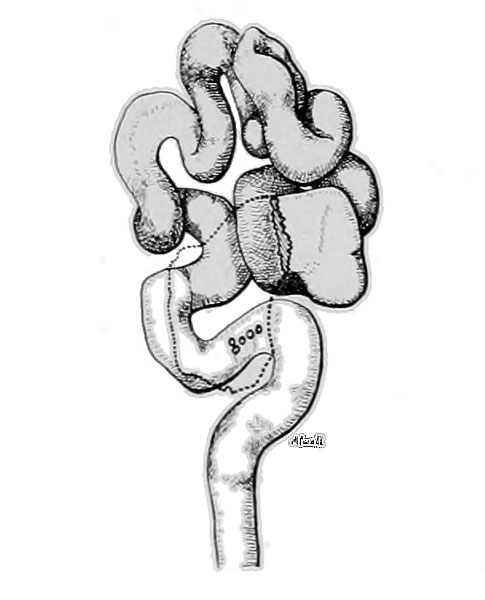
Fig. 9 Model of the left oviduct of rat
Model of the left oviduct of rat Xo. 62, 2 days, 22 hours. X 10. Not quite the entire oviduct was available for reconstruction, thus the relative position of the upper end of the uterine horn is not shown in this figure. Fimbriated end and infundibulum removed in the drawing, so as to expose the underlying loops; their relative position is given in dotted outline. The position of five 2 cell stages, found within this tube, is given as if seen through a transparent wall.
| Historic Disclaimer - information about historic embryology pages |
|---|
 |
- Albino Rat Links: Fig 14. Right Oviduct | Fig 15. 8 and 11-cell stages | The Development of the Albino Rat 1915
Cite this page: Hill, M.A. (2024, May 19) Embryology Huber1915 1fig09.jpg. Retrieved from https://embryology.med.unsw.edu.au/embryology/index.php/File:Huber1915_1fig09.jpg
- © Dr Mark Hill 2024, UNSW Embryology ISBN: 978 0 7334 2609 4 - UNSW CRICOS Provider Code No. 00098G
| Historic Disclaimer - information about historic embryology pages |
|---|
|
Cite this page: Hill, M.A. (2024, May 19) Embryology Huber1915 1fig09.jpg. Retrieved from https://embryology.med.unsw.edu.au/embryology/index.php/File:Huber1915_1fig09.jpg
- © Dr Mark Hill 2024, UNSW Embryology ISBN: 978 0 7334 2609 4 - UNSW CRICOS Provider Code No. 00098G
File history
Click on a date/time to view the file as it appeared at that time.
| Date/Time | Thumbnail | Dimensions | User | Comment | |
|---|---|---|---|---|---|
| current | 11:06, 7 April 2013 | | 485 × 600 (34 KB) | Z8600021 (talk | contribs) | {{Huber1915 figures}} {{Huber1915_footer}} |
You cannot overwrite this file.
File usage
The following 3 pages use this file: