File:Hertig1946b fig08.jpg

Original file (800 × 652 pixels, file size: 134 KB, MIME type: image/jpeg)
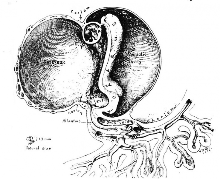
Fig. 8. A drawing of the right half of a 7 somite (1.7 mm.) embryo of the 4th week of development
At this stage, the blood vessels in the embryo, body stalk and chorion are anatomically complete although not quite so in the yolk-sac. The yolk-sac at its junction with the embryo is beginning to eonstrict and is associated with the invagination of the gut entoderm to form the fore and hind gut. (Fig. 2 from Cullen’s “The Umbilicus and Its Diseases,” W. B. Saunders Company.)
References
Hertig AT. lnvolution of tissues in fetal life: a review. (1946) Anat. Rec. 94: 96-116.
Cite this page: Hill, M.A. (2024, May 11) Embryology Hertig1946b fig08.jpg. Retrieved from https://embryology.med.unsw.edu.au/embryology/index.php/File:Hertig1946b_fig08.jpg
- © Dr Mark Hill 2024, UNSW Embryology ISBN: 978 0 7334 2609 4 - UNSW CRICOS Provider Code No. 00098G
File history
Click on a date/time to view the file as it appeared at that time.
| Date/Time | Thumbnail | Dimensions | User | Comment | |
|---|---|---|---|---|---|
| current | 17:17, 7 August 2017 | | 800 × 652 (134 KB) | Z8600021 (talk | contribs) |
You cannot overwrite this file.
File usage
The following page uses this file: