File:Hertig1946b fig07b.jpg
Original file (800 × 1,068 pixels, file size: 108 KB, MIME type: image/jpeg)
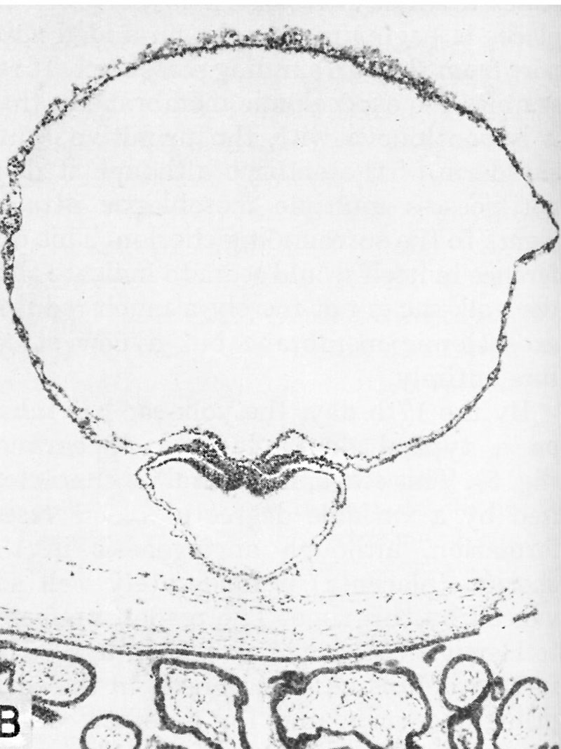
Fig. 7.B. A human embryo of approximately 20 - 21 days of age
A cross section through the middle of a 20 - 21 day embryo. Note the enormous size of the yolk-sac containing blood islands Within its doublelayered wall. The embryo proper is the trilaminar mass between the yolk-sac and amniotic cavity and consists, from within outward, of primitive or gut entoderm, mesoderm and ectoderm. A portion of the chorion is seen below. Carnegie 7545, section 6-2-2, X60.
References
Hertig AT. lnvolution of tissues in fetal life: a review. (1946) Anat. Rec. 94: 96-116.
Cite this page: Hill, M.A. (2024, May 11) Embryology Hertig1946b fig07b.jpg. Retrieved from https://embryology.med.unsw.edu.au/embryology/index.php/File:Hertig1946b_fig07b.jpg
- © Dr Mark Hill 2024, UNSW Embryology ISBN: 978 0 7334 2609 4 - UNSW CRICOS Provider Code No. 00098G
File history
Click on a date/time to view the file as it appeared at that time.
| Date/Time | Thumbnail | Dimensions | User | Comment | |
|---|---|---|---|---|---|
| current | 17:09, 7 August 2017 | | 800 × 1,068 (108 KB) | Z8600021 (talk | contribs) |
You cannot overwrite this file.
File usage
The following page uses this file: