File:HamiltonBoyd1960 plate01.jpg

Original file (1,000 × 1,021 pixels, file size: 350 KB, MIME type: image/jpeg)
Plate 1
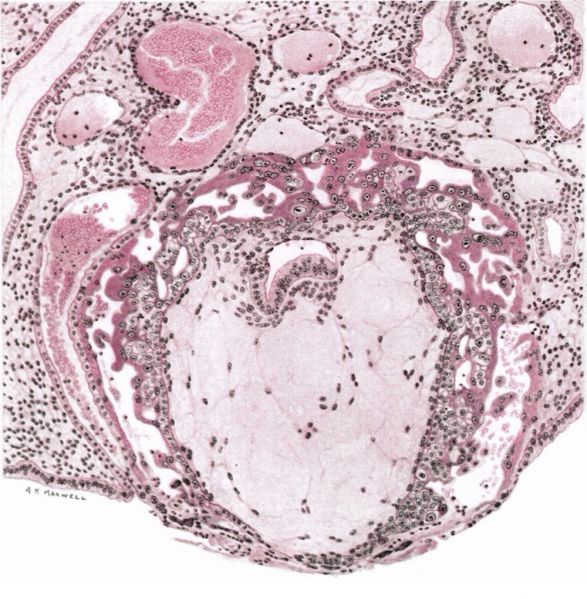
Fig. 1. A drawing ( x 140) of a section through the middle of an implantation site at the 11th-12th day of development (Barnes embryo).
The surface view of the implantation site is shown in Pl. 2, fig. 2.
The blastocystic trophoblast has differentiated into primitive syncytium and cytotrophoblast ; the latter surrounds the primitive mesoblast which almost completely fills the © original blastocyst cavity. At intervals the cytotrophoblast has proliferated to form projections which are the forerunners of the ‘primary villi’. The primitive syncytium, which has not yet extended completely round the superficial aspect of the implantation site, shows an extensive development of intercommunicating lacunae containing some maternal blood cells. The endometrium surrounding the implanted ovum is oedematous and there is haemorrhage into a uterine gland in contact with the syncytium.
Photomicrographs of selected portions of the trophoblast of this specimen are illustrated in Pl. 4, figs. 9-12.
Plates: 1 | 2 | 3 | 4 | 5 | 6 | 7 | 8 | 9 | 10 | 11 | 12 | 13
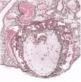
Plate 1
Plate 2
Plate 3
Plate 4
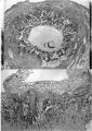
Plate 5
Plate 6
Plate 7
Plate 8
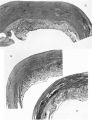
Plate 9
Plate 10
Plate 11
Plate 12
Plate 13


Reference
Hamilton WJ. and Boyd JD. Development of the human placenta in the first three months of gestation. (1960) J Anat. 94(3): 297-328. PMID14399291 | PDF
Cite this page: Hill, M.A. (2024, May 8) Embryology HamiltonBoyd1960 plate01.jpg. Retrieved from https://embryology.med.unsw.edu.au/embryology/index.php/File:HamiltonBoyd1960_plate01.jpg
- © Dr Mark Hill 2024, UNSW Embryology ISBN: 978 0 7334 2609 4 - UNSW CRICOS Provider Code No. 00098G
File history
Click on a date/time to view the file as it appeared at that time.
| Date/Time | Thumbnail | Dimensions | User | Comment | |
|---|---|---|---|---|---|
| current | 12:21, 6 August 2020 | | 1,000 × 1,021 (350 KB) | Z8600021 (talk | contribs) | crop, adjust size |
| 12:20, 6 August 2020 | 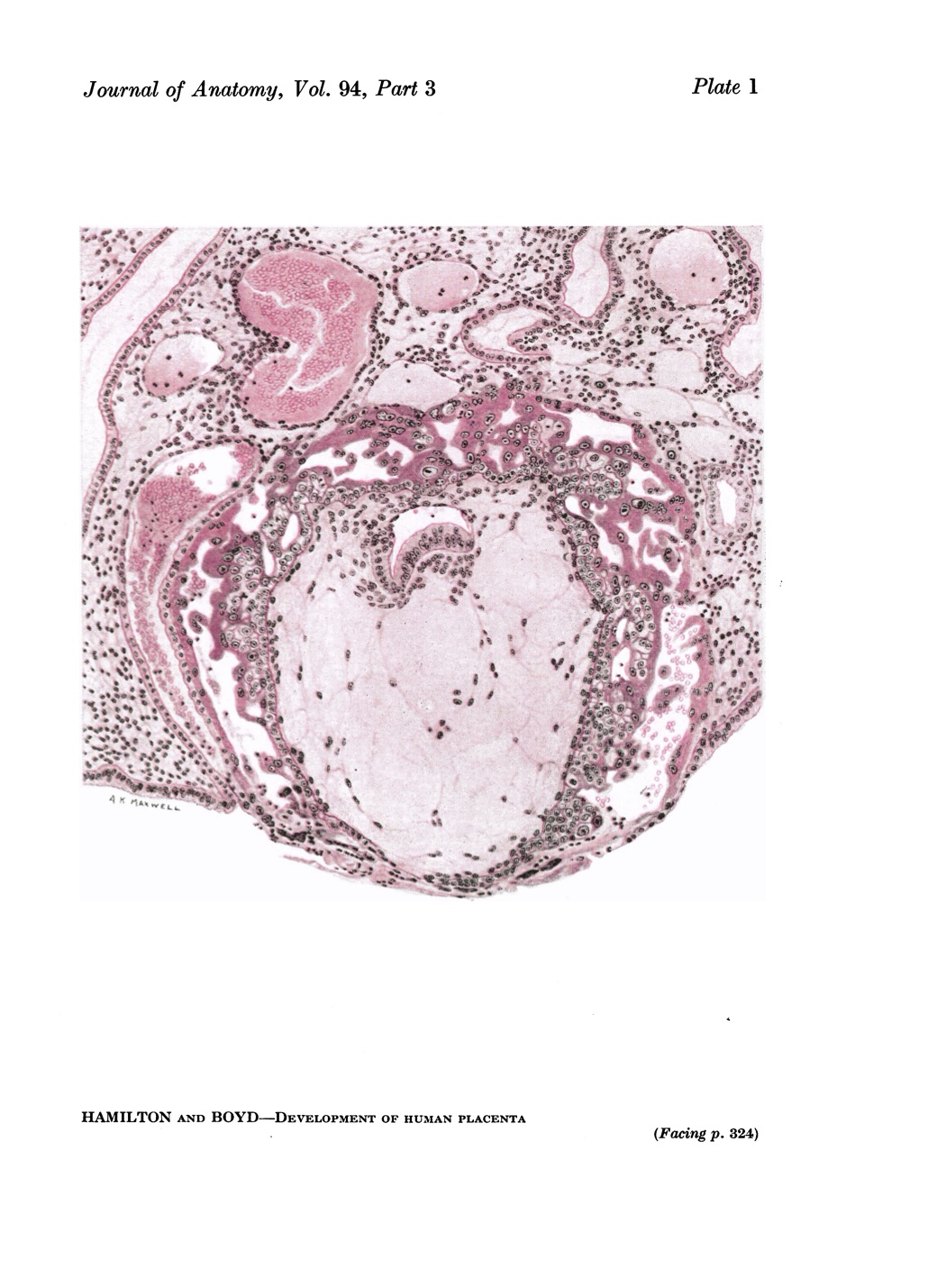 | 1,030 × 1,429 (284 KB) | Z8600021 (talk | contribs) |
You cannot overwrite this file.
File usage
The following 12 pages use this file:
- Paper - Development of the human placenta in the first three months of gestation (1960)
- File:HamiltonBoyd1960 fig02.jpg
- File:HamiltonBoyd1960 fig03.jpg
- File:HamiltonBoyd1960 fig04.jpg
- File:HamiltonBoyd1960 fig05.jpg
- File:HamiltonBoyd1960 fig06.jpg
- File:HamiltonBoyd1960 fig07.jpg
- File:HamiltonBoyd1960 fig08.jpg
- File:HamiltonBoyd1960 plate02.jpg
- File:HamiltonBoyd1960 plate03.jpg
- File:HamiltonBoyd1960 plate13.jpg
- Template:HamiltonBoyd1960 plates