File:Gray1160.jpg
Gray1160.jpg (600 × 549 pixels, file size: 72 KB, MIME type: image/jpeg)
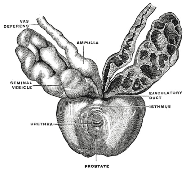
Prostate Gland
Prostate with seminal vesicles and seminal ducts, viewed from in front and above. (Spalteholz.)
The prostate (Fig. 1160) is a firm, partly glandular and partly muscular body, which is placed immediately below the internal urethral orifice and around the commencement of the urethra. It is situated in the pelvic cavity, below the lower part of the symphysis pubis, above the superior fascia of the urogenital diaphragm, and in front of the rectum, through which it may be distinctly felt, especially when enlarged. It is about the size of a chestnut and somewhat conical in shape, and presents for examination a base, an apex, an anterior, a posterior and two lateral surfaces.
The base (basis prostatæ) is directed upward, and is applied to the inferior surface of the bladder, The greater part of this surface is directly continuous with the bladder wall; the urethra penetrates it nearer its anterior than its posterior border.
The apex (apex prostatæ) is directed downward, and is in contact with the superior fascia of the urogenital diaphragm.
- Prostate Links: Image - Prostate Gland | Image - Prostate and Seminal Vesicles | Image - Bladder and Seminal Vesicles | Prostate Development | Male Development
- Gray's Images: Development | Lymphatic | Neural | Vision | Hearing | Somatosensory | Integumentary | Respiratory | Gastrointestinal | Urogenital | Endocrine | Surface Anatomy | iBook | Historic Disclaimer
| Historic Disclaimer - information about historic embryology pages |
|---|
 |
| iBook - Gray's Embryology | |
|---|---|

|
|
Reference
Gray H. Anatomy of the human body. (1918) Philadelphia: Lea & Febiger.
Cite this page: Hill, M.A. (2024, June 1) Embryology Gray1160.jpg. Retrieved from https://embryology.med.unsw.edu.au/embryology/index.php/File:Gray1160.jpg
- © Dr Mark Hill 2024, UNSW Embryology ISBN: 978 0 7334 2609 4 - UNSW CRICOS Provider Code No. 00098G
File history
Click on a date/time to view the file as it appeared at that time.
| Date/Time | Thumbnail | Dimensions | User | Comment | |
|---|---|---|---|---|---|
| current | 12:27, 28 October 2010 | | 600 × 549 (72 KB) | S8600021 (talk | contribs) | ==Prostate Gland== Prostate with seminal vesicles and seminal ducts, viewed from in front and above. (Spalteholz.) {{Template:Gray}} Gray's 1918 Anatomy |
You cannot overwrite this file.
File usage
The following 3 pages use this file: