File:Gray0908.jpg
Gray0908.jpg (500 × 359 pixels, file size: 30 KB, MIME type: image/jpeg)
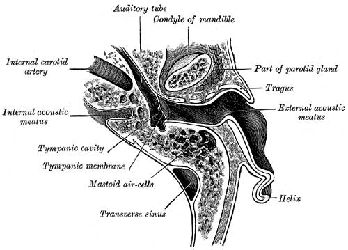
Horizontal Section through Left Ear
Horizontal section through left ear; upper half of section.
The External Acoustic Meatus
(meatus acusticus externus; external auditory canal or meatus) extends from the bottom of the concha to the tympanic membrane (Fig. 907, Fig. 908). It is about 4 cm. in length if measured from the tragus; from the bottom of the concha its length is about 2.5 cm. It forms an S-shaped curve, and is directed at first inward, forward, and slightly upward (pars externa); it then passes inward and backward (pars media), and lastly is carried inward, forward, and slightly downward (pars interna). It is an oval cylindrical canal, the greatest diameter being directed downward and backward at the external orifice, but nearly horizontally at the inner end. It presents two constrictions, one near the inner end of the cartilaginous portion, and another, the isthmus, in the osseous portion, about 2 cm. from the bottom of the concha. The tympanic membrane, which closes the inner end of the meatus, is obliquely directed; in consequence of this the floor and anterior wall of the meatus are longer than the roof and posterior wall.
(Text modified from Gray's 1918 Anatomy)
- Gray's Images: Development | Lymphatic | Neural | Vision | Hearing | Somatosensory | Integumentary | Respiratory | Gastrointestinal | Urogenital | Endocrine | Surface Anatomy | iBook | Historic Disclaimer
| Historic Disclaimer - information about historic embryology pages |
|---|
 |
| iBook - Gray's Embryology | |
|---|---|

|
|
Reference
Gray H. Anatomy of the human body. (1918) Philadelphia: Lea & Febiger.
Cite this page: Hill, M.A. (2024, May 23) Embryology Gray0908.jpg. Retrieved from https://embryology.med.unsw.edu.au/embryology/index.php/File:Gray0908.jpg
- © Dr Mark Hill 2024, UNSW Embryology ISBN: 978 0 7334 2609 4 - UNSW CRICOS Provider Code No. 00098G
File history
Click on a date/time to view the file as it appeared at that time.
| Date/Time | Thumbnail | Dimensions | User | Comment | |
|---|---|---|---|---|---|
| current | 00:19, 28 September 2009 | | 500 × 359 (30 KB) | S8600021 (talk | contribs) |
You cannot overwrite this file.
File usage
The following 4 pages use this file: