File:Gray0032.jpg
From Embryology
Size of this preview: 719 × 599 pixels.
Original file (800 × 667 pixels, file size: 159 KB, MIME type: image/jpeg)
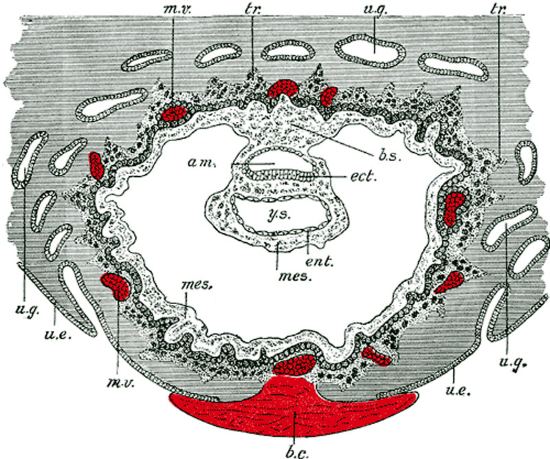
Human Embryo Week 3
Original label was "Day 8 to 9" though showing the trilaminar embryo suggests late week 2 to early week 3 of development. Uterine cavity is shown at bottom of image.
Section through ovum imbedded in the uterine decidua. Semidiagrammatic. (After Peters.)
- am. - amniotic cavity
- b.c. - blood clot, at the site of initial implantation
- b.s. - body-stalk, or connective stalk later forming the placental cord region with placental blood vessels
- ect. - embryonic ectoderm that will contribute to embryonic and placental membrane development
- ent. - entoderm (endoderm), this was the historic term for what we today call endoderm that will contribute to embryo development
- mes. - mesoderm, consisting of both embryonic mesoderm (in the trilaminar embryonic disc) and extraembryonic mesoderm (outside the trilaminar embryonic disc)
- m.v. - maternal vessels, spiral arteries that have been opened at their ends
- tr. - trophoblast, relative to the embryonic disc the outer syncitiotrophoblast and inner cytotrophoblast layers that will contribute to placental development
- u.e. - uterine epithelium, the epithelial layer that lines the unerus
- u.g. - uterine glands, the glands that secrete nutrients to support the initial growth both before and after implantation
- y.s. - yolk-sac, the endoderm lined and extraembryonic mesoderm covered cavity that will contribute to the gastrointestinal tract, blood and blood vessels
- Links: Implantation | Week 3 |
- Gray's Images: Development | Lymphatic | Neural | Vision | Hearing | Somatosensory | Integumentary | Respiratory | Gastrointestinal | Urogenital | Endocrine | Surface Anatomy | iBook | Historic Disclaimer
| Historic Disclaimer - information about historic embryology pages |
|---|
 |
| iBook - Gray's Embryology | |
|---|---|

|
|
Reference
Gray H. Anatomy of the human body. (1918) Philadelphia: Lea & Febiger.
Cite this page: Hill, M.A. (2024, May 13) Embryology Gray0032.jpg. Retrieved from https://embryology.med.unsw.edu.au/embryology/index.php/File:Gray0032.jpg
- © Dr Mark Hill 2024, UNSW Embryology ISBN: 978 0 7334 2609 4 - UNSW CRICOS Provider Code No. 00098G
File history
Click on a date/time to view the file as it appeared at that time.
| Date/Time | Thumbnail | Dimensions | User | Comment | |
|---|---|---|---|---|---|
| current | 11:12, 22 May 2011 | | 800 × 667 (159 KB) | MarkHill (talk | contribs) | ==Human Embryo Day 8 to 9== Section through ovum imbedded in the uterine decidua. Semidiagrammatic. (After Peters.) original figure title * '''am.''' - amniotic cavity * '''b.c.''' - blood clot, at the site of initial implantation * '''b.s.''' - body-st |
You cannot overwrite this file.
File usage
The following 17 pages use this file:
- 2009 Lecture 8
- 2010 Lecture 8
- ANAT2341 Lab 4 - Implantation and Villi Development
- ASA Meeting 2013 - Placenta
- Anatomy of the Human Body by Henry Gray
- BGDA Practical 3 - Extraembryonic Spaces
- BGDA Practical 3 - Implantation
- BGDA Practical 3 - Week 2 Summary
- BGDA Practical Placenta - Implantation and Early Placentation
- Foundations Practical - Week 1 to 8
- Implantation
- Lecture - Placenta Development
- Placenta - Cord
- Placenta - Membranes
- Placenta Development
- Yolk Sac Development
- Talk:ANAT2341 Lab 4 - Implantation and Villi Development