File:Frazer1915 fig05.jpg

Original file (1,000 × 529 pixels, file size: 104 KB, MIME type: image/jpeg)
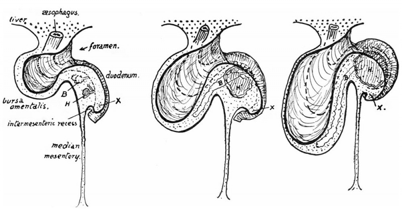
Fig. 5. Schemes to illustrate the formation of the curve of the duodenum
The mesentery is supposed to be viewed from the dorsum, having been cut away from its dorsal attachment. The dorsal mesoduodenum is seen as a thick mass projecting to the right immediately below the protrusion to the left of the small sac. The alimentary tube is shown by interrupted lines running down in the front wall of the small sac, turning to the right, and becoming continuous with the duodenum which passes down in front of the mesoduodenum.
The head (H) and body (B) of pancreas grow into the mesoduodenum: observe that B can pass almost directly into the wall of the small sac. The other figures show how, while B extends into this wall, H grows in the mesoduodenum and by its enlargement curves out the duodenum round it: thus the duodenum is attached all the time to the mesoduodenum. X is the position of Treitz’ band, put in its hypothetical place in the first two figures.
| Historic Disclaimer - information about historic embryology pages |
|---|
 |
- Links: Fig 1 | Fig 2 | Fig 3 | Fig 4 | Fig 5 | Fig 6 | Fig 7 | Fig 8 | Fig 9 | Fig 10 | Fig 11 | Fig 12 | Fig 13 | Fig 14 | Fig 15 | Fig 16 | Fig 17 | Fig 18 | 1915 Frazer | Intestine Development | Category:Intestine
Reference
Frazer JE. and Robbins RH. On the factors concerned in causing rotation of the intestine in man. (1915) J Anat. 50(1): 75-110. PMID 17233053
Cite this page: Hill, M.A. (2024, April 27) Embryology Frazer1915 fig05.jpg. Retrieved from https://embryology.med.unsw.edu.au/embryology/index.php/File:Frazer1915_fig05.jpg
- © Dr Mark Hill 2024, UNSW Embryology ISBN: 978 0 7334 2609 4 - UNSW CRICOS Provider Code No. 00098G
File history
Click on a date/time to view the file as it appeared at that time.
| Date/Time | Thumbnail | Dimensions | User | Comment | |
|---|---|---|---|---|---|
| current | 11:37, 7 January 2017 | | 1,000 × 529 (104 KB) | Z8600021 (talk | contribs) | |
| 11:35, 7 January 2017 | 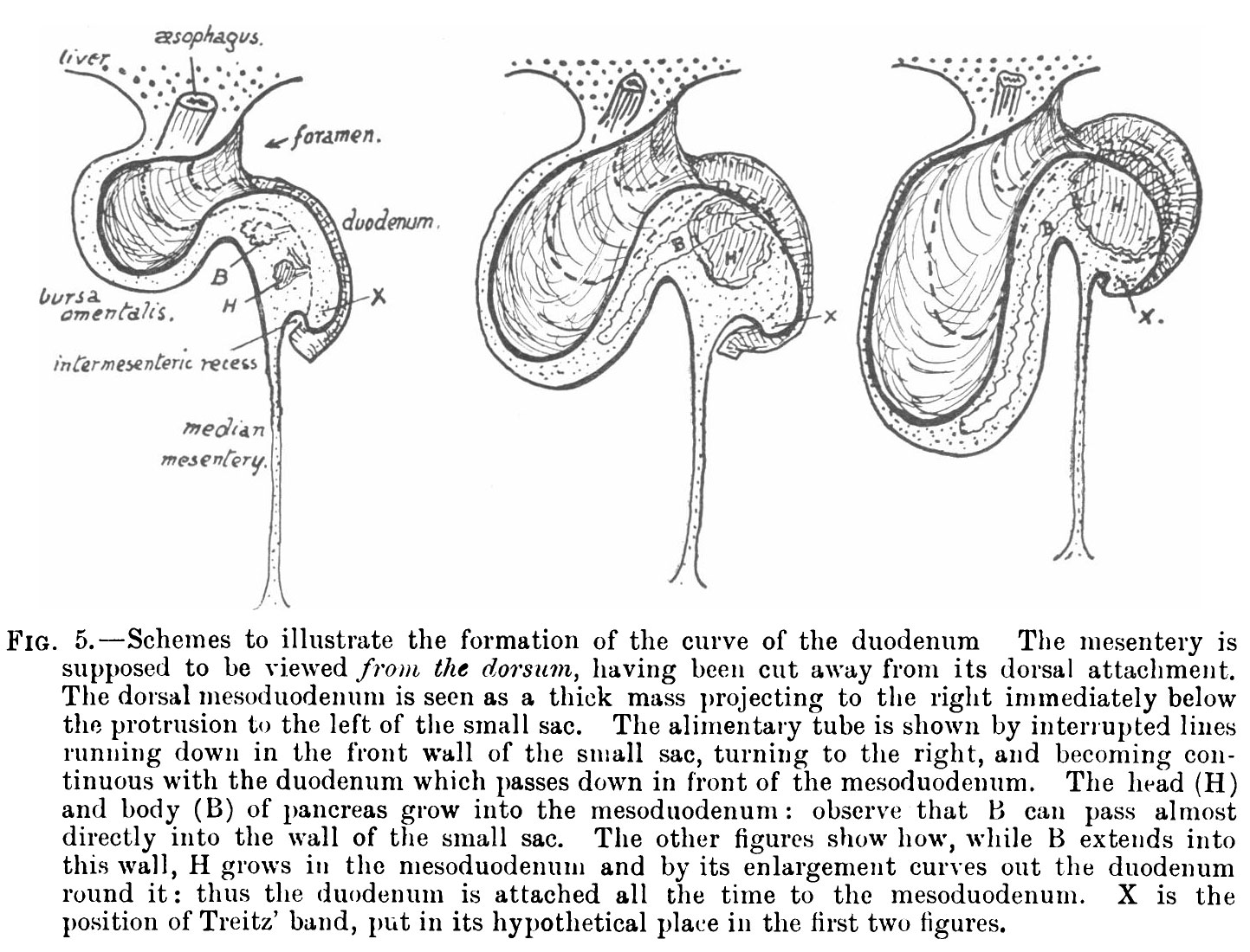 | 1,443 × 1,100 (330 KB) | Z8600021 (talk | contribs) |
You cannot overwrite this file.
File usage
The following 4 pages use this file: