File:Frazer1911 fig01.jpg
From Embryology
Size of this preview: 800 × 479 pixels. Other resolution: 1,280 × 766 pixels.
Original file (1,280 × 766 pixels, file size: 187 KB, MIME type: image/jpeg)
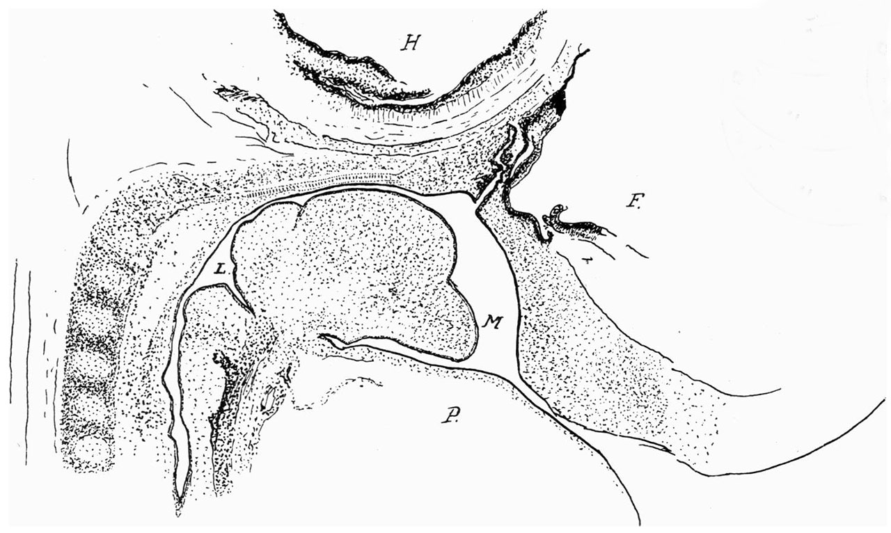
Fig. 1. Sagittal section 12 mm Embryo
(camera lucida). H., hind-brain; F., fore-brain ; P., pericardium ; M., mouth (placed in front of mandibular arch) ; L., upper part of opening larynx.
| Historic Disclaimer - information about historic embryology pages |
|---|
 |
Reference
Frazer JE. Pharyngeal end of Rathke's pouch. (1911) J Anat. 45: 190-196. PMID 17232879
Cite this page: Hill, M.A. (2024, May 8) Embryology Frazer1911 fig01.jpg. Retrieved from https://embryology.med.unsw.edu.au/embryology/index.php/File:Frazer1911_fig01.jpg
- © Dr Mark Hill 2024, UNSW Embryology ISBN: 978 0 7334 2609 4 - UNSW CRICOS Provider Code No. 00098G
File history
Click on a date/time to view the file as it appeared at that time.
| Date/Time | Thumbnail | Dimensions | User | Comment | |
|---|---|---|---|---|---|
| current | 10:13, 9 January 2017 | | 1,280 × 766 (187 KB) | Z8600021 (talk | contribs) | |
| 10:12, 9 January 2017 | 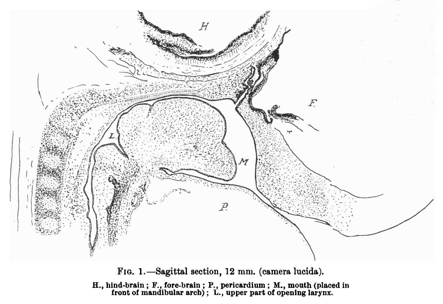 | 1,537 × 1,053 (246 KB) | Z8600021 (talk | contribs) |
You cannot overwrite this file.
File usage
The following 2 pages use this file: