File:Fetal ovary meiosis 03.jpg
Fetal_ovary_meiosis_03.jpg (652 × 400 pixels, file size: 64 KB, MIME type: image/jpeg)
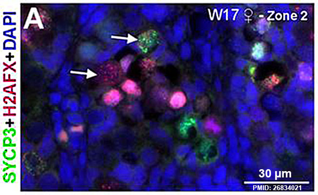
Human Fetal Ovary Meiosis (second trimester)
(A) High magnifications of histological sections of a human ovary at week 15 (GA week 17) immunostained for the meiotic markers H2AFX (red) and SYCP3 (green) showing zone 2. A/Ai - image | full image
White arrows point to germ cells showing decondensed chromatin and various degrees of H2AFX and SYCP3 expression.
Note that primordial follicles (indicated by white asterisks), characteristic of zone 3, were negative for both H2AFX and SYCP3. Ai,Bi,Ci,Di, DAPI channel only.
Scale bars are 30 µm.
Reference
Heeren AM, He N, de Souza AF, Goercharn-Ramlal A, van Iperen L, Roost MS, Gomes Fernandes MM, van der Westerlaken LA & Chuva de Sousa Lopes SM. (2016). On the development of extragonadal and gonadal human germ cells. Biol Open , 5, 185-94. PMID: 26834021 DOI.
Copyright
© 2016. Published by The Company of Biologists Ltd This is an Open Access article distributed under the terms of the Creative Commons Attribution License (http://creativecommons.org/licenses/by/3.0), which permits unrestricted use, distribution and reproduction in any medium provided that the original work is properly attributed.
Panel A figure 6 cropped, sharpened, resized and relabelled.
Cite this page: Hill, M.A. (2024, May 2) Embryology Fetal ovary meiosis 03.jpg. Retrieved from https://embryology.med.unsw.edu.au/embryology/index.php/File:Fetal_ovary_meiosis_03.jpg
- © Dr Mark Hill 2024, UNSW Embryology ISBN: 978 0 7334 2609 4 - UNSW CRICOS Provider Code No. 00098G
File history
Click on a date/time to view the file as it appeared at that time.
| Date/Time | Thumbnail | Dimensions | User | Comment | |
|---|---|---|---|---|---|
| current | 09:47, 21 May 2016 | | 652 × 400 (64 KB) | Z8600021 (talk | contribs) |
You cannot overwrite this file.
File usage
The following page uses this file: