File:Fetal membrane and placenta cartoon.jpg
Fetal_membrane_and_placenta_cartoon.jpg (600 × 429 pixels, file size: 125 KB, MIME type: image/jpeg)
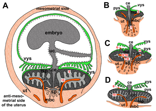
Schematic view of the Guinea Pig Fetal Membranes and the Placenta
A The schematic view demonstrates the arrangement of the mesometrially situated embryo, the chorioallantoic placenta, positioned at the antimesometrial side, and the other fetal membranes inside the uterus, representing an advanced stage of pregnancy.
B In initial pregnancy, the placenta contains mainly of trophospongium with the developing subplacenta confluent to the main placenta. Strings of extraplacental trophoblast and syncytial streamers can be followed from towards the maternal blood channels.
C In early pregnancy, a labyrinth is established and the subplacenta represents a distinct organ.
D Near term, the placenta is highly villous with the labyrinth as the dominant area. The subplacenta has been reduced.
Legend
- Red numbers in white boxes refer to subsequent figures with more detailed documentation of specific regions.
- Ce = central excavation
- lab = labyrinth
- mbc = maternal blood channel
- pys = parietal yolk sac
- sp = subplacenta
- sys = syncytial streamers
- tsp = trophospongium
- uc = umbilical cord
- ut = uterine tissue
- vys = visceral yolk sac
Reference
<pubmed>18771596</pubmed>| PMCID: PMC2543018
This is an Open Access article distributed under the terms of the Creative Commons Attribution License (http://creativecommons.org/licenses/by/2.0), which permits unrestricted use, distribution, and reproduction in any medium, provided the original work is properly cited.
File history
Click on a date/time to view the file as it appeared at that time.
| Date/Time | Thumbnail | Dimensions | User | Comment | |
|---|---|---|---|---|---|
| current | 09:21, 16 August 2009 | | 600 × 429 (125 KB) | S8600021 (talk | contribs) | Schematic view of the fetal membranes and the placenta. (A) The schematic view demonstrates the arrangement of the mesometrially situated embryo, the chorioallantoic placenta, positioned at the antimesometrial side, and the other fetal membranes inside |
You cannot overwrite this file.
File usage
The following 4 pages use this file: