File:Enbom1939 fig01.jpg
Original file (1,250 × 572 pixels, file size: 55 KB, MIME type: image/jpeg)
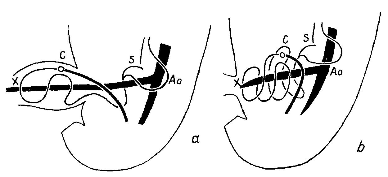
Fig. 1a and b. Diagrammatic side View of plastiline models of opossum embryos (a) 9.4 mm and (b) 11.2 mm
Linear course of the intestinal canal with the omphalomesenteric artery axial in the C-onvolute system. In the older foetus the pliysiological umbilical hernia has been drawn in as a consequence of the vitelline artery having been shortened by one—half, the gastro-enterie coil in its entirety then being pressed together with dorsal tlisplacexnent. A0 = aorta together with a. omphalomesenteriea.
C = coecum. S = stomach. ac = point at which vitelline artery leaves mesentery. Large intestine drawn with thicker line. 8/1.
Reference
Enbom G. The early looping of the alimentary canal in the mammalian and human foetus and the mechanisms assumed to be active in this process. (1939) Anat. Rec. 75(3): 409-413.
Cite this page: Hill, M.A. (2024, June 1) Embryology Enbom1939 fig01.jpg. Retrieved from https://embryology.med.unsw.edu.au/embryology/index.php/File:Enbom1939_fig01.jpg
- © Dr Mark Hill 2024, UNSW Embryology ISBN: 978 0 7334 2609 4 - UNSW CRICOS Provider Code No. 00098G
File history
Click on a date/time to view the file as it appeared at that time.
| Date/Time | Thumbnail | Dimensions | User | Comment | |
|---|---|---|---|---|---|
| current | 14:05, 16 February 2018 | | 1,250 × 572 (55 KB) | Z8600021 (talk | contribs) | |
| 14:04, 16 February 2018 |  | 1,353 × 977 (155 KB) | Z8600021 (talk | contribs) | '''Fig.1a and b''' Diagrammatic side View of plastiline models of opossum embryos, (:1) 9.4 mm. and (b) 11.2 mm. Linear course of the intestinal canal with the omphalomesenteric artery axial in the C-onvolute system. In the older foetus the pliysiologi... |
You cannot overwrite this file.
File usage
The following page uses this file: