File:Corner1920 fig01.jpg
Original file (1,000 × 606 pixels, file size: 159 KB, MIME type: image/jpeg)
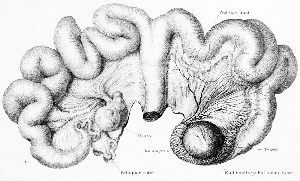
Figure 1 General view of Abnormal Pig Ovary, Testis and Uterus
X 0.75.
The uterus was normally formed (fig. 1), presenting two cornua as usual. Its size corresponded to that of the uterus of a young, sexually mature sow. On the right side the uterine cornu ended in a normal Fallopian tube in connection with a normal ovary; the latter contained four recent corpora lutea, one of them cystically dilated. On washing out the contents of the tube with saline solution, one ovum was found, normal in all respects except that the cytoplasm was slightly shrunken; one polar body had been extruded. The left uterine horn, normal in size and form, ended in a very slender tube about 1 mm. in external diameter near the uterus, which gradually thinned down to an almost linear dimension, losing its lumen, and finally ending in the connective tissue over the epididymis (fig. 1).
On the left side, in place of an ovary there was a mass 30 by 25 by 20 mm. in diameter, of dull flesh-color, exactly resembling a testis in form, texture, and color. It was covered by a thick capsule in which large and somewhat tortuous vessels coursed; when this tunic was incised the contents bulged over the cut edges. The exposed surface was dry and granular in appearance.
On this side of the uterus there was a well-defined Wolffian duct, such as is occasionally present in sows, beginning in the vagina and running parallel to the uterine horn between the layers of the broad ligament. However, instead of ending in a cul-de-sac or in a series of minute cysts in the region of the ovarian pedicle, as this duct usually does when present in the sow, it became greatly convoluted as it approached the tip of the cornu and finally so closely coiled as to form the body indicated in figure 1. This structure presented the appearance of an epididymis by reason of its texture, its close apposition to the testis-like body, and also because of a slight constriction at the middle portion, suggesting a division into globus major and minor.
| Historic Disclaimer - information about historic embryology pages |
|---|
 |
Glossary Links
- Glossary: A | B | C | D | E | F | G | H | I | J | K | L | M | N | O | P | Q | R | S | T | U | V | W | X | Y | Z | Numbers | Symbols | Term Link
Cite this page: Hill, M.A. (2024, May 8) Embryology Corner1920 fig01.jpg. Retrieved from https://embryology.med.unsw.edu.au/embryology/index.php/File:Corner1920_fig01.jpg
- © Dr Mark Hill 2024, UNSW Embryology ISBN: 978 0 7334 2609 4 - UNSW CRICOS Provider Code No. 00098G
File history
Click on a date/time to view the file as it appeared at that time.
| Date/Time | Thumbnail | Dimensions | User | Comment | |
|---|---|---|---|---|---|
| current | 07:35, 27 December 2012 | | 1,000 × 606 (159 KB) | Z8600021 (talk | contribs) |
You cannot overwrite this file.
File usage
The following page uses this file: