File:Cooper1938 plate29.jpg

Original file (1,200 × 2,329 pixels, file size: 359 KB, MIME type: image/jpeg)
Plate XXIX
Fig. 11. “ Septum " between two tertiary bronchial branchings. Cap. = capillary; R.B.C. = erythrocyte. 18 cm. embryo. X650.
Fig. 12. Primary, secondaries and tertiaries. Approaching full term. X 80.
Fig. 13. Tertiary branchings showing continuous lining. A detached cell is seen at A. Cap. = capillary; R.B.C. = erythrocyte. Approaching full term. X450.
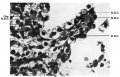
Fig. 11.
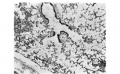
Fig. 12.
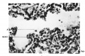
Fig. 13.


| Historic Disclaimer - information about historic embryology pages |
|---|
 |
Reference
Cooper ERA. A histological investigation of the development and structure of the human lung. (1938) J Pathology 47: 105-114.
Cite this page: Hill, M.A. (2024, May 11) Embryology Cooper1938 plate29.jpg. Retrieved from https://embryology.med.unsw.edu.au/embryology/index.php/File:Cooper1938_plate29.jpg
- © Dr Mark Hill 2024, UNSW Embryology ISBN: 978 0 7334 2609 4 - UNSW CRICOS Provider Code No. 00098G
File history
Click on a date/time to view the file as it appeared at that time.
| Date/Time | Thumbnail | Dimensions | User | Comment | |
|---|---|---|---|---|---|
| current | 13:15, 27 November 2016 | | 1,200 × 2,329 (359 KB) | Z8600021 (talk | contribs) | |
| 13:15, 27 November 2016 |  | 1,367 × 2,653 (423 KB) | Z8600021 (talk | contribs) |
You cannot overwrite this file.
File usage
The following page uses this file: