File:Congdon1922-17.jpg

Original file (1,000 × 411 pixels, file size: 55 KB, MIME type: image/jpeg)
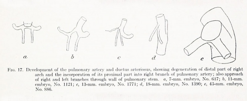
Fig. 17. Development of the pulmonary artery and ductus arteriosus
Showing degeneration of distal part of right arch and the incorporation of its proximal part into right branch of pulmonary artery.
Also approach of right and left branches through wall of pulmonary stem.
- a 7-mm. embryo, No. 617
- b 11-mm. embryo, No. 1121
- c 13-mm. embryo, No. 1771
- d 18-mm. embryo, No. 1390
- e 43-mm. embryo, No. 886
- Human Aortic Arch 1922: Table 1 | Fig. 1-16 | Fig 17 | Fig 18-25 | Fig 18 | Fig 19 | Fig 20 | Fig 21 | Fig 22 | Fig 23 | Fig 24 | Fig 25 | Fig 26 | Fig 27-28 | Fig 29 | Fig 30 | Fig 31 | Fig 32 | Fig 33 | Fig 34 | Fig 35 | Fig 36 | Fig 37 | Fig 38 | Fig 39 | Fig 40 | Plate 1 | Plate 2 | Plate 3 | Carnegie No.68 | Volume XIV | Contributions to Embryology | Historic Disclaimer | Cardiovascular Development | Respiratory Development
Reference
Congdon ED. Transformation of the aortic-arch system during the development of the human embryo. (1922) Contrib. Embryol., Carnegie Inst. Wash. Publ 277, 14:47-110.
Cite this page: Hill, M.A. (2024, May 11) Embryology Congdon1922-17.jpg. Retrieved from https://embryology.med.unsw.edu.au/embryology/index.php/File:Congdon1922-17.jpg
- © Dr Mark Hill 2024, UNSW Embryology ISBN: 978 0 7334 2609 4 - UNSW CRICOS Provider Code No. 00098G
| Historic Disclaimer - information about historic embryology pages |
|---|
 |
File history
Click on a date/time to view the file as it appeared at that time.
| Date/Time | Thumbnail | Dimensions | User | Comment | |
|---|---|---|---|---|---|
| current | 15:30, 1 September 2012 | 1,000 × 411 (55 KB) | Z8600021 (talk | contribs) | ||
| 17:38, 7 May 2011 | 1,144 × 428 (64 KB) | S8600021 (talk | contribs) | ==Fig. 17. Development of the pulmonary artery and ductus arteriosus== Showing degeneration of distal part of right arch and the incorporation of its proximal part into right branch of pulmonary artery; also approach of right and left branches through wa |
You cannot overwrite this file.
File usage
The following page uses this file: