File:CPAMXCT.jpg

Original file (1,559 × 838 pixels, file size: 157 KB, MIME type: image/jpeg)
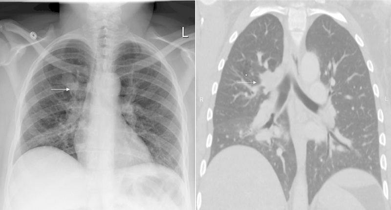
Congenital Pulmonary Airway Malformation
Diagnostic imaging of a 36-year old woman diagnosed with CPAM. On the left shows the chest radiography with the arrow pointing to the lobulated medial right upper lobe nodule. The photo on right shows a contrast CT image with another arrow in pointing to the same lobulated medial right upper lobe nodules as seen in the left image, however the CT revealed surrounding cystic hyperlucency.
References
<pubmed>PMC4821328</pubmed>
Copyright © 2015 The Authors This is an open access article under the CC BY-NC-ND license (http://creativecommons.org/licenses/by-nc-nd/4.0/).
- Note - This image was originally uploaded as part of an undergraduate science student project and may contain inaccuracies in either description or acknowledgements. Students have been advised in writing concerning the reuse of content and may accidentally have misunderstood the original terms of use. If image reuse on this non-commercial educational site infringes your existing copyright, please contact the site editor for immediate removal.
File history
Click on a date/time to view the file as it appeared at that time.
| Date/Time | Thumbnail | Dimensions | User | Comment | |
|---|---|---|---|---|---|
| current | 01:38, 5 October 2017 | | 1,559 × 838 (157 KB) | Z5178462 (talk | contribs) | ==Congenital Pulmonary Airway Malformation== Diagnostic imaging of a 36-year old woman diagnosed with CPAM. On the left shows the chest radiography with the arrow pointing to the lobulated medial right upper lobe nodule. The photo on right shows a con... |
You cannot overwrite this file.
File usage
The following file is a duplicate of this file (more details):
The following 2 pages use this file: