File:Bremer1906 fig08.jpg
From Embryology
Size of this preview: 800 × 399 pixels. Other resolution: 1,154 × 576 pixels.
Original file (1,154 × 576 pixels, file size: 124 KB, MIME type: image/jpeg)
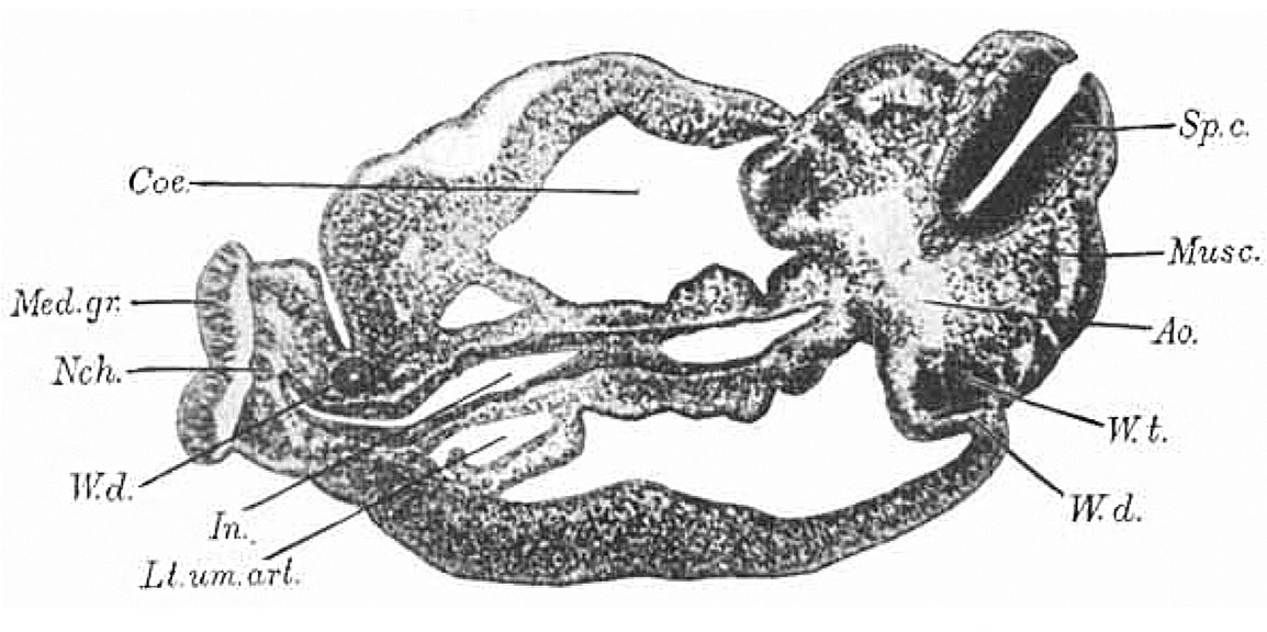
Fig.8. Section through body of embryo, near the cloaca
Section through body of Harvard Embryo Template:HE714, near the cloaca. x 60.
| Historic Disclaimer - information about historic embryology pages |
|---|
 |
- Human Embryo 4 mm: Fig. 1. Brain and Pharynx Models | Fig. 2-5. Head Sections | Fig. 6. Brain Model | Fig. 7. Embryo Drawing | Fig. 8. Body Section | Fig. 9. Heart Model
Reference
Bremer JL. Description of a 4-mm human embryo. (1906) Amer. J Anat. 5: 459-480.
Cite this page: Hill, M.A. (2024, May 15) Embryology Bremer1906 fig08.jpg. Retrieved from https://embryology.med.unsw.edu.au/embryology/index.php/File:Bremer1906_fig08.jpg
- © Dr Mark Hill 2024, UNSW Embryology ISBN: 978 0 7334 2609 4 - UNSW CRICOS Provider Code No. 00098G
File history
Click on a date/time to view the file as it appeared at that time.
| Date/Time | Thumbnail | Dimensions | User | Comment | |
|---|---|---|---|---|---|
| current | 18:48, 16 July 2015 | | 1,154 × 576 (124 KB) | Z8600021 (talk | contribs) | |
| 18:47, 16 July 2015 | 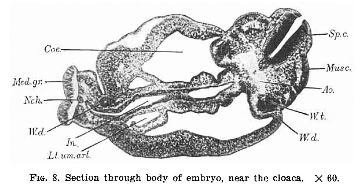 | 1,200 × 637 (138 KB) | Z8600021 (talk | contribs) | {{Bremer1906 figures}} |
You cannot overwrite this file.
File usage
The following 2 pages use this file: